To get started:
- Click the edit this page tab at the top. Save the page after each step, then edit it again.
- Click the 3D button (when editing, above the wikitext box) to insert Jmol.
- show the Scene authoring tools, create a molecular scene, and save it. Copy the green link into the page.
- Add a description of your scene. Use the buttons above the wikitext box for bold, italics, links, headlines, etc.
More help: Help:Editing
For more help, look at this link:
http://www.proteopedia.org/wiki/index.php/Help:Getting_Started_in_Proteopedia
Syntaxin-1A
Introduction
Syntaxin-1A is part of the SNARE (soluble N-ethylmaleimide-sensitive factor attachment protein receptor) family. It is an integrin plasma membrane protein found almost exclusively in neurons and it is known for its essential roles in neuronal signaling. [1] It assembles in tight core complexes, which promote fusion of carrier vesicles with target compartments. Members of the SNARE class of proteins are expressed in all eukaryotic cells and are distributed in distinct subcellular compartments.
The Williams syndrome is caused by the deletion of genetic material from a specific region of chromosome 7. The deleted region includes more than 25 genes, and researchers believe that a loss of several of these genes contributes to the characteristic features of this disease. The deleted region has now been identified and syntaxin-1A is one of the genes that has been deleted.[2]
Structure
Syntaxin-1A has 288 residues. Residue 1-265 make up the cytoplasmic domain and residue 266-288 form the carboxyl-terminal transmembrane anchor. It adapts a well-folded tertiary structure with repeated forming a colied coil structure.[3]
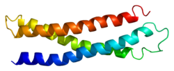
NMR spectroscopy was used to identify a folded N-terminal domain in Syntaxin-1A and to determine its three-dimensional structure. This 120 residue N-terminal domain is conserved in plasma membrane syntaxins. The domain is made up from three long and an up-and-down bundle with a left-handed twist. Two are displayed within the SNARE complex.[4]
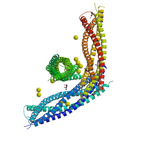
The image below shows a neuronal synaptic fusion complex to exemplify the use of the , ASP 68. SNARE proteins are involved in the fusion of vesicles with their target membranes but the structural details of these complexes are still unknown. X-ray is used here to show the crystal structure at 2.4 Å resolution of a core synaptic fusion complex containing syntaxin-1A, synaptobrevin-II and SNAP-25B.[5]
Mechanism of action
Syntaxin-1A has an essential role in neuronal signaling. It regulates fusion, docking and trafficking of synaptic vesicles with the plasma membrane.
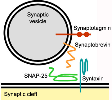
The SNARE complex is a four-alpha-helix bundle composed of syntaxin-1A, synaptobrevin and SNAP-25. In neuronal exocytosis, syntaxin and synaptobrevin are attached in respective membranes by their C-terminal domains, whereas SNAP-25 is connected to the plasma membrane via several cysteine-linked palmitoyl chains. Synaptotagmin acts as a calcium sensor and regulates the SNARE closure. The influx of calcium into the cell triggers the completion of the assembly reaction.
The protein also plays an important role in internalization of vesicles containing a transporter for the neurotransmitter glutamate. The transportation of glutamate is important to avoid excessive stimulation of glutamate receptors and to support the conversion to the inhibitory neurotransmitter GABA. Syntaxin-1A also regulates ion channels such as Ca2+ channels, K+ channels and epithelial Na+ channels.[6]
Applications
As mentioned before, the neurodevelopmental disorder Williams syndrome is related to the deletion of, among others, syntaxin-1A. Other typical genes that are deleted are CLIP2, ELN, GTF2I, GTF2IRD1, and LIMK1. The most common symptoms of Williams syndrome are mental disability, heart defects, and unusual facial features.[7]
Noradrenaline transporters (NETs) terminate noradrenergic transmission and are often used as a target for antidepressant medications. Syntaxin-1A interacts with NET and modulates NET activity. Protein kinase C activation disrupts NET/syntaxin-1A interactions and down regulates NET activity. Syntaxin-1A binds the NH2 terminal of NET. The protein both supports the surface trafficking of NET and limits the transporter function through its direct interaction with NET. The net result of these two factors is a dynamic cycle of interactions that leads to coordinated control between noradrenaline release and uptake.[8]
References
- ↑ Yu, Yong-Xin, Li Shen, Peng Xia, et al. "Syntaxin 1A promotes the endocytic sorting of EAAC1 leading to inhibition of glutamate transport." 119.18 (2006): 3776-3787. Web. 25 April. 2012. <http://stke.sciencemag.org/cgi/content/full/joces;119/18/3776>
- ↑ Rowe, J, F Calegari, R Longhi, and P Rosa. "Syntaxin 1A is delivered to the apical and basolateral domains of epithelial cells: the role of munc-18 proteins."PubMed. 114.18 (2001): 3323-32. Web. 26 April. 2012. <http://www.ncbi.nlm.nih.gov/pubmed/11591820>
- ↑ Lerman, Jeffrey, James Robblee, Robert Fairman, et al. "Structural Analysis of the Neuronal SNARE Protein Syntaxin-1A." Biochemistry. 39.29 (2000): 8470–8479. Web. 1 May. 2012. <http://pubs.acs.org/doi/abs/10.1021/bi0003994>
- ↑ Fernandez, I, J Ubach, I Dulubova, et al. "Three-dimensional structure of an evolutionarily conserved N-terminal domain of syntaxin 1A."Cell(Cambridge,Mass.). 94. (1998): 841-849. Web. 2 May. 2012. <http://www.rcsb.org/pdb/explore/explore.do?structureId=1BR0>
- ↑ Sutton, R.B., D. Fasshauer, R. Jahn, et al. "Crystal structure of a SNARE complex involved in synaptic exocytosis at 2.4 A resolution." Nature. 395 (1998): 347-353. Web. 26 May. 2012. <http://www.rcsb.org/pdb/explore/explore.do?structureId=1sfc>
- ↑ Ray, Brian. "Endocytotic Function for Syntaxin 1A."Science. 2006.353 (2006): tw322. Web. 27 May. 2012. <http://stke.sciencemag.org/cgi/content/abstract/sigtrans;2006/353/tw322>
- ↑ Rowe, J, F Calegari, R Longhi, and P Rosa. "Syntaxin 1A is delivered to the apical and basolateral domains of epithelial cells: the role of munc-18 proteins."PubMed. 114.18 (2001): 3323-32. Web. 26 April. 2012. <http://www.ncbi.nlm.nih.gov/pubmed/11591820>
- ↑ Sung, Uhna, Aurelio Galli, Kristopher Kahlig, et al. "A Regulated Interaction of Syntaxin 1A with the Antidepressant-Sensitive Norepinephrine Transporter Establishes Catecholamine Clearance Capacity." Journal of Neuroscience. 23.5 (2003): 1697-1709. Web. 28 May. 2012. <http://www.jneurosci.org/content/23/5/1697.full>
|


