Sandbox Reserved 963
From Proteopedia
Anti-amyloid-beta Fab WO2 (Form A, P212121)
Alzheimer's disease (AD), is the most common form of dementia. It is a chronic neurodegenerative disease that usually develops slowly and get worse over time, becoming severe enough to interfere with daily tasks. The most common early symptom of Alzheimer's disease is the difficulty in remembering recently learned information. As the patient with Alzheimer’s disease ages, symptoms such as speaking problems, language problems, mood swings, disorientation, behavioural issues, and loss of motivation, can appear. Acetylcholinesterase inhibitors such as rivastigmine, tacrine, donepezil and galantamine or, memantine, which is a NMDA receptor antagonist, are used to treat the patients suffering from AD but unfortunately, the benefit from their use is small. It is important to understand that none of these medications stops the disease itself. However, many groups of researchers are seeking a solution to this problem and most of them are currently focused on the activity of a small peptide called Amyloid β (Aβ). One of the hypothetical causes of this disease is depicted as the presence of the amyloid plaques (which are composed of Aβ) found in the brains of Alzheimer patients. These are peptides of 36–43 amino acids, obtained via proteolysis of Amyloid Precursor Protein (APP) by β and γ secretases. An immunologic approach to the disease is made. Researchers have developped a monoclonal antibody, WO2, which can bind specifically to the Aβ’s epitope, thus leading the complex to be phagocyted.
Structural highlightsThe three-dimensional structure of WO2 was obtained thanks to X-ray crystallography. The structure of WO2 that is described here is that of WO2 murine anti-Aβ monoclonal Fab (antigen binding fragment) in the space group P212121 (Form A). There's also another form available for the WO2 antibody, the Form B, but it is not mentioned here because it is crystallized in the space group P21. Thus, in what follows, the different features correspond to the Form A. This structure appears to be that of a typical immunoglobulin (Ig) Fab heavy-chain/light-chain heterodimer weighting 52362 Da[1]. The heavy chain is made up of 252 amino acids while the light chain is made up of 224 amino acids.[2]
WO2 contains several helices and sheets[3] :
Constant (C) and variable (V) domains of both light (L) and heavy (H) chains include the following residues : VL(1-107), CL(108-212), VH(2-113) and CH(114-213). represent a loop in the sequence between Complementarity Determining Regions (CDR) heavy-chain 1 (H1) and H2, located in the hinge region of the Fab away from the ligand binding site. However, due to the poor electron density on this loop, some uncertainties about the accuracy of the model in this region can be found. is a non-CDR loop involved in symmetry-related close contacts. of the light chain is the only residue to stay outside from allowed regions of the Ramachandran plot. The unfavorable φ / Ψ torsion angles arise from the fact that this residue is in a γ-turn restrained by the (i to i+2) hydrogen bond between .[6] In the WO2 Form A structure, were found, involved in crystal contacts, and bound to Asp1L of WO2.
Interactions with the ligand Aβ1-16Overview of the WO2:Aβ1-16 complexAβ1-16 represents the minimal zinc binding domain and contains the entire immunodominant B-cell epitope of Aβ, it is therefore interesting to see how this fragment of Aβ interacts with WO2. The main residues which closely contact the CDRs of WO2 by sitting within the antigen binding site of WO2 are Ala2 to Ser8 and they stretch 20 Å from the N-terminus to the C-terminus.
The surface area of the Aβ2-8 structure is 1118 Ų, from which 60% is buried (665 Ų) in the antibody interface. In addition, we note two significant interfaces between Aβ and WO2 : 367 Ų of its surface contacts the heavy chain and 298 Ų contacts the light chain. We notice that residues in the middle of the Aβ1-16 structure exhibit lower B-factors than atoms at the N- and C- terminus of the Aβ1-16 peptide, indicating that they are more flexible (since the B-factor, also called the temperature factor, represents the relative vibrational motion of different parts of a structure and atoms with low B-factors belong to a part of the structure quite rigid whereas atoms with high B-factors generally belong to part of a structure that is very flexible[1]). Phe4 and His6 are completely buried in the Fab interface, each with nearly half of their surface area buried in the VH interface and half in the VL interface. All other residues are located exclusively at the interface with either the VH or the VL domains. Residues of the light chain closely contacting Aβ residues include , and from light-chain CDR 1 (L1) and . All residues from Phe4 to Ser8, except Asp7, make close contact with the WO2 heavy-chain CDRs. Close contacting interface residues include and . Also, we observe no contact between Aβ and the L2 or H1 CDRs of WO2 and there is no evidence in the structure of any water-mediated contacts between WO2 and Aβ.[7]
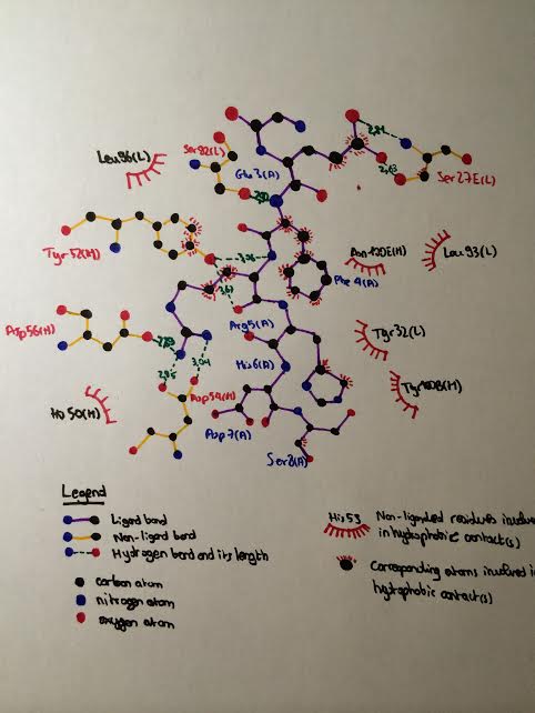
Details of the close interactions between WO2 and Aβ2-8As mentionned previously, the residues of Aβ closely interacting with the CDRs of WO2 extend from Ala2 to Ser8. Let's focus on the interactions of each of these residues with the antibody (see the following LIGPLOT scheme) : Interactions with Ala2Ala2 of Aβ2-8 is recognized by WO2 through a hydrogen bond between its main chain carbonyl group and the amide group of . Interactions with Glu3Hydrogen bond between the side chain of Glu3 and the main-chain amide of . Additionally, there are potential salt bridges between the side chain of Glu3 and Nδ1/Nε2 atoms of . Interactions with Phe4Hydrogen bond between the amide group of Phe4 and the carbonyl group of Ser92L. Interactions with Arg5Arg5 is a donor for 5 side chain-side chain hydrogen bonds and 1 main chain-side chain hydrogen bond, involving , and as shown below :
Interactions with His6His6 contacts both heavy- and light-chain elements with 10 side chain-side chain hydrophobic contacts with . Van der Waals contacts have been detected between His6 and of WO2. With Phe4 and Arg5, His6 represents the core of the epitope of Aβ which sits at the heavy-chain/light-chain junction of the CDRs of WO2. Interactions with Ser8Ser8 makes a single Van der Waals contact with of WO2. The special case of Asp7Asp7 sits above a large water-filled cavity in the Fab antigen binding site but makes no direct or water-mediated contacts with WO2.
Comparison of unliganded and liganded WO2 Fab structuresUnliganded and liganded structures superimpose very closely with an r.m.s.d. (root-mean-square deviation) of 0.3 Å on all Cα atoms (the r.m.s.d. is the measure of the average distance between the atoms of superimposed proteins[2]). Even the CDRs of liganded and unliganded states are barely distinguishable. Except for some small variations (<1 Å) around (L1), , and , there is no substantial change in the CDRs when Aβ binds with WO2. Moreover, thanks to temperature-factors analysis, it appears that CDR H1 is less flexible in the liganded structure.[8]
Biological RelevanceOne of the most common isoforms of Aβ is the 42-mer Aβ (with the sequence : 1-DAEFRHDSGYEVHHQKLVFFAEDVGSNKGAIIGLMVGGVVIA-42), which is the most fibrillogenic isoforms and is, therefore linked to disease states. Aβ42 damages and kills neurons by generating reactive oxygen species when it self-aggregates. Aβ self-aggregation is promoted by its binding with metal ions (such as Cu2+, Zn2+, etc) by reason of, among others, its His6, His13, His14, Tyr10, Asp1 and Glu11 residues. If this process occurs on the neurons' membrane, it causes lipid peroxidation and the generation of a toxic aldehyde called 4-hydroxynonenal which, in return, impairs the function of ion-motive ATPases, glucose transporters and glutamate transporters. It also triggers depolarization of the synaptic membrane, excessive calcium influx and mitochondrial impairment, making neurons vulnerable to excitotoxicity and apoptosis : this is known as the beginning of the neurodegenerative process of AD.[9] The important role of metals in AD is highlighted by a metal chelator, the clioquinol, which reduces amyloid plaques in the brain of AD patients. The mAb (monoclonal antibody) WO2 recognises the N-terminus of Aβ. This region of Aβ constitutes the immunodominant B-cell epitope of Aβ and lacks T-cell epitopes involved in the toxicity of previous clinical trials. Some experiments showed scientists that the great interest of WO2 lies in the fact that when it binds Aβ, it prevents Asp1 and His6 of Aβ to participate in metal coordination, preventing Aβ from aggregating and thus, neutralizing harmful effects of Aβ.[10] Related Structures
ContributorsKerim Secener and Nicolas Pautrieux References
| ||||||||||||

