Introduction
The neurotensin receptor (NTSR1) belongs to the superfamily of proteins known as G protein-coupled receptors (GPCRs) and responds to the 13 amino acid hormone neurotensin (NTS). Currently around 800 G protein-coupled receptors have been identified and are hypothesized to be responsible for roughly 80% of signal transduction.[1] GPCRs are involved in a vast array of physiological processes within the body that range from interactions with dopamine to effects on secretion of bile in the intestines.[2] [3] Due to the vast array of functions that these proteins serve and their high abundance within the body, these proteins have become major drug targets.[4]
A critical topic in the understanding of GPCRs is the transition from the inactive to active state. This transition is responsible for the transduction of a signal from the extracellular to the intracellular space. The transition occurs when a ligand, NTS in the case of NTSR1, binds to the receptor causing a conformational change in the protein that leads to the activation of the intracellular G protein. Currently no crystal structures of the receptor in its unbound, inactive form exist making the transition difficult to study. [5] NTSR1 can be seen in blue and the ligand NTS can be seen in green.
Neurotensin
Neurotensin (NTS) is a 13-amino acid peptide originally isolated from bovine hypothalamus. [6] NTS fulfills the roles of both a neurotransmitter and a neuromodulator in the nervous system and a hormone in the periphery nervous system. NTS is a neuromodulator of dopamine transmission and of anterior pituitary hormone secretion. [7] In the periphery of the digestive tract and cardiovascular system, NTS is a paracrine and endocrine modulator.[8] Finally, NTS serves as a growth factor for many normal and cancerous cell types. [9]
Only the C-terminal tail of NTS, amino acids 8-13, were resolved in the .(PDB code:4GRV) Amino acids 8-13 are only resolved because these are the amino acids that interact with NTRS1 to produce the conformational change that is responsible for G-protein activation. The peptide is colored by element: carbon, oxygen, nitrogen.
Structure
Overall Structure
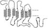
Figure 1: Neurotensin Incorporation in Membrane. This image depicts the spanning of the membrane made by NTSR1 and illustrates the need for transduction from its extracellular binding site to the intracellular region. (PubMed).
Like other G protein-coupled receptors, the neurotensin receptor is composed of 3 distinct regions. An where neurotensin binds and causes a conformational change of the protein. A region containing (PDB code:4GRV) that transduce the signal from the extracellular side of the cell membrane to the intracellular side. Lastly, an intracellular region, that when activated by a conformational change in the protein, activates a G-protein associated with this receptor. Currently no crystal structures of the inactive form of the neurotensin receptor are available. Without a representation of the inactive form, the conformational changes caused by agonist binding are still not completely known.
Neurotensin Binding Site
Binding of NTS to the binding site on NTSR1 is enriched by (PDB code:4GRV)between the positive NTS arginine side chains and the electronegative pocket. The protein is colored by charge: negative and positive. Two of NTS's arginine residues are colored blue. In addition, the C-terminus of NTS forms a (PDB code:4GRV) with Arg328 of NTSR1. Three hydrogen bonds are made between the side chains of NTS and the receptor while most of the interactions are a result of Van der Waals interactions. The binding pocket is partially capped by a at the proximal end of the receptor's N-terminus.[5] The interactions in the binding site cause a wide spread conformational change in the receptor leading to the receptor to adopt an active conformation. This active conformation allows for the activation of the associated intracellular G-protein.
Hydrophobic Stacking
A major player in the transduction of the extracellular signal to the intracellular G-protein is the (PDB code:4GRV)that links the bound hormone with the hydrophobic core of the neurotensin receptor. The carboxylate of Leu13 of NTS forms a hydrogen bond network with R327, R328, and Y324. The Tyr324, in turn, is brought into an orientation to make the formation of a (PDB Code:4XEE) network between F358, W321, A157, and F317 possible.[10] The effects that this network has on the activation of the intracellular G-protein was examined by the mutagenesis of amino acids that disrupted the formation of this network. Mutagenesis of , , , , , and (PDB code:4GRV) showed that when this interaction was disrupted, the receptor no longer was able to activate the G-protein. [10] This discovery lead to the conclusion that the conformational changes caused by this stacking allows for the signal to be moved from the extracellular binding site through the transmembrane helices of the receptor to the intracellular region activating the G-protein.
Sodium Binding Pocket
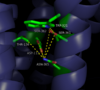
Figure 2: Closed form of sodium binding pocket that caps the entrance of sodium into the top of the binding pocket. (PDB Code:
4XEE)
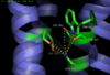
Figure 3: Open form of sodium binding pocket that does not cap the entrance of sodium into the top of the binding pocket. (PDB code:
4GRV)
Conserved across all class A GPCRs, a (PDB code:
4GRV) is seen in the middle of TM2 helix. The sodium ion is coordinated with a highly conserved Asp113 and four other oxygen contacts from a combination of water molecules. For G-protein activation to be possible, a hydrogen bond coordination with T156, S362, and N365 of the NPxxY
motif must occur. Trp321 helps to maintain the active conformation of the receptor by occluding the top of the binding pocket using
Van der Waals interactions (Figure 2). This occlusion stops sodium ions from entering the top of the binding pocket and helps NTSR1 remain in its active conformation. The conformation of the binding pocket where Trp321 does not occlude the top can be seen when mutations to A86L, G215A, and V360A are present (Figure 3). This form of the receptor would allow more sodium into the binding pocket and therefor stabilize the inactive receptor form.
[11]
Allosteric Effects
Sodium ions are a negative allosteric inhibitor to the binding of the neurotensin agonist to the binding site on the neurotensin receptor. Sodium's binding causes for the receptor to favor its inactive state by disrupting the hydrogen bond network between nearby amino acids. This disruption in the hydrogen bond network causes the pocket to be in its uncollapsed form. Asp113 of the highly conserved D/RY motif and Asn365 of the highly conserved NPxxY motif form a substantial hydrogen bonding network with T156 and S362.[10] This hydrogen bonding network prevents the incorporation of the sodium ion by collapsing upon itself and filling the sodium binding pocket. Trp321 also works to inhibit the incorporation of the sodium ion by capping off the sodium binding pocket to not allow sodium to enter from the top. Trp321 uses Van der Waals interactions to place it in the conformation necessary to block sodium from entering the site. By not allowing for sodium to enter this binding site, the receptor is able to conform to its active state and activate the G-protein that is associated with it.
Clinical Relevance
NTSR1 is commonly expressed in various invasive
cancer cell lines making it a promising cancer drug target. It is prevalent in
colon cancer adenocarcinoma, but is not found in adult colon cell types.
[12] NTSR1 is also found in aggressive
prostate cancer cells, but not
epithelial prostate cells. In prostate cancer cells, binding of NTS results in
mitogen-activated protein kinase (PKB),
phosphoinositide-3 kinase (PI-3K),
epidermal growth factor receptor (EGFR),
SRC, and
STAT5 phosphorylation.
[12] These all result in increased DNA synthesis,
cell proliferation, and survival. Inhibition of NTSR1 and its downstream signaling represents a target for
radiotherapy, which uses radiation to target malignant cells.
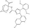
Figure 4: Meclinerant: An inhibitor of NTSR1 found to enhance selectivity of radiotherapy in cancer treatment (PubMed).
NTSR1 can be inhibited by agonist
meclinertant which inhibits proliferation and prosurvival of cancer cells. Combination treatment of radiation and meclinerant provides selective treatment of cancer cells over normal cells, indicating the need for clinical trials of this approach.
[13]


