User:Ashley R. Wilkinson/Sandbox 1
From Proteopedia
Contents |
Metabotropic Glutamate Receptor mGluR-2
Introduction
Metabotropic glutamate receptorsmGluR2 Background are found in the central nervous system and play a critical role in modulating cell excitability and synaptic transmission (Lin). Glutamate is the main neurotransmitter in the brain and activates 8 different types of metabotropic glutamate receptors (Seven). Metabotropic Glutamate Receptor 2 (mGlu2) is a member of the Class C GPCR Family and can further be classified into the Group II subgroup of metabotropic receptors. Since mGlu2 is a part of the Class C GPCR family, it undergoes small conformational changes to the transmembrane domain (TMD) to move from the inactive to the fully active structure (Shuling). Functionality of mGlu2 will be dependent on the concentration of glutamate. Higher concentrations of glutamate will promote stronger signal transduction from the extracellular domain to the transmembrane domain.
mGlu2 plays vital roles in memory formation, pain management, and addiction, which makes it an important drug target for Parkinson’s Disease (Zhang), Schizophrenia (blue link), Cocaine Addiction (Yang) and many other neurological conditions.
Overview of Structure
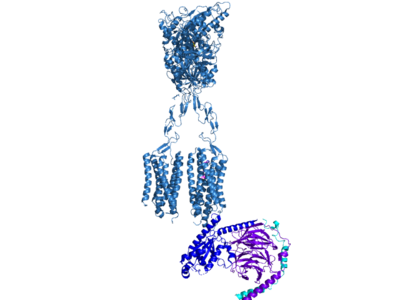
Cryo-EM studies of mGlu2 have yielded adequate structures that have acted as maps to aid in producing a better structural understanding of the inactive and active states of mGlu2 (Lin). The overall structure of the mGlu2 is composed of 3 main parts: a ligand binding Venus FlyTrap Domain(VFT), followed by a Cysteine Rich Domain linker to the Transmembrane Domain that contains 7 alpha helices (7TM) on both the that aid in the binding of the G-Protein. Class C CPCRs such as mGlu2, are activated by their ability to form dimers. MGlu2 is a homodimer which is imperative to the receptor’s ability to relay signals induced by glutamate from the extracellular domain(ECD) to its transmembrane domain(TMD). The homodimer of mGlu2 contains an alpha chain and a beta chain. Occupation of both ECDs with the agonist, glutamate, is necessary for a fully active mGlu2. However, only one chain in the dimer is responsible for activation of the G-protein, this suggests an asymmetrical signal transduction mechanism for mGlu2.
|
This is a sentence about the structure of my protein. This is a sentence about the structure of my protein. This is a sentence about the structure of my protein. This is a sentence about the structure of my protein. This is a sentence about the structure of my protein. This is a sentence about the structure of my protein.
Function
Disease
Relevance
Structural highlights
This is my new image.This is a sample scene created with SAT to by Group, and another to make of the protein. You can make your own scenes on SAT starting from scratch or loading and editing one of these sample scenes.
</StructureSection>
References
<7mts/> Du, Juan, et al. “Structures of Human mglu2 and mglu7 Homo- and Heterodimers.” Nature News, Nature Publishing Group, 16 June 2021, https://www.nature.com/articles/s41586-021-03641-w. Lin, Shuling, et al. “Structures of GI-Bound Metabotropic Glutamate Receptors mglu2 and mglu4.” Nature News, Nature Publishing Group, 16 June 2021, https://www.nature.com/articles/s41586-021-03495-2. “Metabotropic Glutamate Receptors.” Abcam, 6 Feb. 2022, https://www.abcam.com/content/metabotropic-glutamate-receptors. Seven, Alpay B., et al. “G-Protein Activation by a Metabotropic Glutamate Receptor.” Nature News, Nature Publishing Group, 30 June 2021, https://www.nature.com/articles/s1586-021-03680-3. Yang, Hong-Ju, et al. “Deletion of Type 2 Metabotropic Glutamate Receptor Decreases Sensitivity to Cocaine Reward in Rats.” Cell Reports, U.S. National Library of Medicine, 11 July 2017, https://www.ncbi.nlm.nih.gov/pmc/articles/PMC5555082/. Zhang, Zhu, et al. “Roles of Glutamate Receptors in Parkinson's Disease.” MDPI, Multidisciplinary Digital Publishing Institute, 6 Sept. 2019, https://dx.doi.org/10.3390%2Fijms20184391.

