Gamma Secretase
Introduction
Background
Gamma Secretase is a transmembrane aspartate protease. It catalyzes peptide bond hydrolysis of type I integral membrane proteins such as Notch, APP, and various other substrates. It recognizes and catalyzes the reaction with its substrate using 3 residue segments. These substrates generate amyloid-β (Aβ). This product is important for various neural processes, and it is well known for its Implications with Alzheimer’s disease (AD). This has made gamma secretase a popular drug target, specifically using gamma secretase (GS) inhibitors. However, due to the nature of gamma secretase having various neural functions, there are dangerous side effects when it is inhibited.
Overall Structure
Γ-secretase is composed of 20 transmembrane components (TMs) and has 4 subunits: Nicastran, Anterior Pharynx-defective 1, Presenilin, and Presenilin Enhancer 2. These subunits are stabilized by hydrophobic interactions and 4 phosphatidylcholines.These phosphatidylcholines have interfaces between: PS1 and PEN-2, APH-1 and PS1, APH-1 and NCT.
Nicastrin (NCT) has a large extracellular domain and 1 TM. It is important to substrate recognition and binding.
Presenilin (PS1) serves as the active site of the protease and contains 9 TMs, each varying in length. The site of autocatalytic cleavage is located between TM6 and TM7 in PS1 and major conformational changes take place in this subunit upon substrate binding.
Anterior pharynx-defective 1 (APH-1) serves as a scaffold for anchoring and supporting the flexible conformational changes of PS1
Activation of the active site is dependent on the binding of Presenilin enhancer 2 (PEN-2). PEN-2 is also important in maturation of the enzyme.
Structural highlights
Substrate Structure
Though gamma secretase has multiple substrates, the substrate of main concern is called Amyloid Precursor Protein (APP). APP is composed of an N-terminal loop, a transmembrane (TM) helix, and a C-terminal β-strand. The uses lateral diffusion as a mechanism of entry into the enzyme, and once in place, the TM helix is anchored by hydrogen bonds. In order to differentiate substrates, the β-strand is often the main point of identification for the enzyme. After this, the helix undergoes unwinding and the process of catalysis can begin.
Lid Complex
Lid is the first point of entry and recognition for the substrate.
Active Site
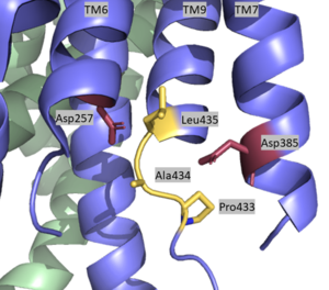
Active Site of Gamma Secretase
The is located between TM6 and TM7 of the PS1 subunit, which is mainly hydrophilic and disordered. Each of these transmembrane helices has an aspartate residue, , which are located approximately 10.6 A˚ apart when inactive.[1] The PAL sequence of is in close proximity with the catalytic aspartates and is important to substrate recognition. Gamma secretase becomes active upon substrate binding, when TM2 and TM6 each rotate about 15 degrees to more closely associate and the two Asp residues hydrogen bond to each other during catalysis. Asp257 and Asp385 are located 6–7 Å away from the scissile peptide bond of the substrate.
Relevance
APP build up leads to Amyloid plaques in brain
Inhibition of γ-secretase could be potential AD treatment
Location of majority of mutations
Over 200 mutations that cause AD
AD mutations heavily target interface between PS1 and APP
Important to look at differences between substrates

