Instructional Information
Figure 1: Bacterial Transformation
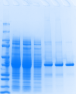
Figure 2: Here is an example of a protein gel. To display a gel, you should be annotate it with size standards and lane numbers.
Adding an uploaded image into your page
1. To add an image of your experiments, you first need to have an image as a JPG or PNG on your computer
2. In your editing page, insert the following code:
[[Image:choose a filename.jpg|thumb|left|250px|Figure insert figure#: insert your caption]]
Uploading the image from your computer
If you are uploading after adding the code to your page
1. Save your page after editing and scroll to the image name on your page. Click on the link.
2. An upload image page should appear.
3. Select the file from your computer that you would like to upload (for example, an annotated gel image or assay figure).
4. Add a summary if you want to
5. Licensing: If this is your own image, select:Own work > "Creative Commons Attribution 3.0
6. Click: Upload File
If you are uploading before adding the code to your page
1. On the "toolbox" menu at the lower left of the screen, click on "Upload File"
2. Next, select the file from your computer that you would like to upload (for example, an annotated gel image or assay figure).
3. Choose a filename that makes sense.
4. Write a summary of your image now, or do this later.
5. Licensing: If this is your own image, select: "Own work" > "Creative Commons Attribution 3.0"
6. Click: Upload File
You can resize your image (change the "250px" to a larger or smaller pixel count), or set the location to right or center as you wish.
Any text you type will show around your thumbnail image, unless you type "clear" within double {{ }}. Take a look at my editing page to see exactly what I have done.
Text Formatting
1. To make your text bold or italic, surround the selected text with 3 or 2 apostrophes (do not use double quotes) on each side, respectively. You can also highlight the text and hit the bold or italics button.
2. To make this cool header, start a new line, insert a space, then type your text
3. To change the color of your text, use the following code: <span style="color:blue;">Insert text here</span> Some colors to try: MediumSeaGreen, SlateBlue, DarkOrchid, Tomato, Navy, Chartreuse or RebeccaPurple. For the full list of color names, see this page.
4. To add a colored background to your text, use the following code: <span style="background:lightgrey;">Insert text here</span>. The same color names as above apply to backgrounds. By combining background and color formatting, you can create text that looks like this
5. For a big "cheat sheet" of formatting, you can click this link
Transcription Factor AphB from V. cholerae (examples of scenes)
Structural highlights
Each AphB comprises an N-terminal (DBD) and a C-terminal (RD). The DBD is connected to the RD via the . AphB subunits are found in two distinct conformations within the tetramer, as determined by the angle between the linker helix and the RD. In the form, the RD and linker helix form an angle of 85 degrees, whereas this angle is 150 degrees in the extended form. Interestingly, the LH forms the dimerization interface between one compact and one extended subunit.
The tetramer (chains A, B, C and D) can be described as a dimer of dimers. Each of two identical dimers are composed of a compact (A and C) and extended (B and D) monomer. The compact and extended subunits differ from one another in the angle between the linker helix and the RD, which is 85° in the compact subunits and 150° in the extended subunits (Fig. 1A). The conformational variability in the AphB subunits can be attributed to the flexible loop connecting the linker helix to the regulatory domain.
The tetramer assembles via two distinct dimerization interfaces (Fig. 2A). The N‐terminal dimerization interface (1142 Å2) is primarily made up of an antiparallel coiled‐coil of the two linker helices, held together by hydrophobic, polar and ionic interactions (Fig. 2A, left inset). The C‐terminal dimerization interface (1414 Å2) forms between regulatory domains, which pack against each other in a head‐to‐tail manner (Fig. 2A, right inset). To form the AphB tetramer, each compact subunit interacts with the regulatory domain of one extended subunit and with the linker helix of the other extended subunit (Fig. 2A and B).


