Background Information
The nucleocapsid protein (or N protein) of SARS-CoV-2 is an important component of the virus particle. This protein's main function is to bind to, pack and stabilize the RNA genome of the virus particle. In this part of the lab, we are going to focus on how a protein can have a structure that facilitates an interaction with another, completely unrelated molecule. In this case, nucleocapsid has a structure that encourages interaction with long RNA molecules (the viral genome) and facilitates the packing of this RNA into the interior of a very small spherical shell.
From a structural point of view, nucleocapsid molecules are also cool because you can clearly see alpha helices, beta-pleated sheets, overall tertiary structure that accommodates RNA interaction and larger quaternary structure that facilitates RNA packing.
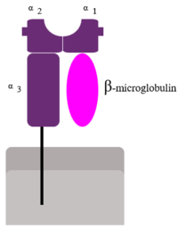
Two subunits of MHC I (alpha--contains alpha 1, 2 and 3 domains--and Beta-microglobulin)
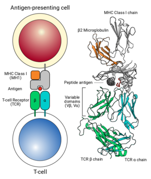
Interaction of MHC Class 1-Antigen complex with TCR
Structure
Take a look at the structure in the box on the right. You'll notice the structure on the right divided into four colors. Each color represents one complete nucleocapsid RNA-binding domain.
1. What do each colored component represent
1. What color is used for the alpha helices?
2. What color is used for the beta sheet?
3. Which subunit(s) (in the 3D model) is/are the MHC molecule? Which subunit(s) is/are the peptide? Think of the hot dog and bun analogy...
4. Which subunit(s) of the MHC molecule play a direct role in binding of the peptide?
- 5. The fit of the peptide in the MHC molecule requires some precision and the peptide must be within a narrow range or lengths. Why do you think this might be (think about the consequences if just any old peptide (of any length) could fit into the MHC groove)?
Hmm...but just how tight does the fit of the peptide look in the MHC groove? Now, open the . Here you are seeing only subunits D and F from the previous image.
6. What is different - or - where did all the space go? To get a feel for this new view and how it relates to the previous model, click on the "popup" button on the bottom right of the structure box. A new window with the MHC 3D structure will open. Then, right click on the 3D image (or ctrl-click for Mac users), then:
a. "Select"-->"all"
b. "Style"-->"structures"-->"backbone"
Now this looks a lot more like the previous MHC model image from parts 1-5.
Click here to reload original:
Surprisingly, the fit of the peptide is not incredibly tight, and many different peptides will fit into any given MHC molecule.
- 7. If you were to alter amino acids in the MHC molecule to (a) affect binding to the peptide, which would you alter? Keep in mind that amino acids within the alpha helices and beta sheets that have side chains that project towards the bound peptide play a crucial role in peptide binding. (b) affect binding to the T cell receptor molecule, which would you alter? (Remember that the T-cell receptor recognizes the whole peptide/binding cleft region as a whole, including amino acids that don’t actually contact the peptide). List 2 amino acids for each answer. (To answer this, move your cursor over an amino acid side chain (light blue) on the image in the area where you would alter something and let the program identify the amino acid for you (three letter code followed by a number at the beginning of the ID string that pops-up).
The following two questions can be answered without the computer. You may want to skip them for now and keep working. Just make sure that you come back to them later!
- 8. What might be the role of the portion of the MHC not involved in direct binding to the peptide? Use your imagination, look at pictures in your book (Chapter 43), or look up and read about MHC on the web (wikipedia would work) and list as many as you can - don’t worry about the true answer, yet!
- 9. A very similar molecule in your immune system is called MHC Class II. (See diagrams in Campbell, Chapter 43 or on wikipedia.) This molecule is structurally and functionally analogous to an MHC Class I molecule. Juvenile diabetes is a disease caused (at least in part) when your MHC Class II molecules erroneously present a peptide from your pancreas. This targets the cells of your pancreas for destruction by your immune system and hence the disease -- called an "autoimmune disease" because you are “immune” to yourself. Interestingly, the cause is a mutation in the genes that code for the MHC molecule. From what you have learned so far, it would be reasonable to assume that the mutation has affected the binding of the MHC to the pancreatic peptide. BUT, while this is in part true, the mutation also disrupts a “salt-bridge” (a weak ionic bond) between the two alpha helices. (An example would be a bridge between Asp77 and His151 in the MHC Class I protein). How could this affect peptide binding? Think back to what you know about how a protein shape is determined.


