is an important protein expressed in several human tissues, such as the heart and kidneys. It plays a key role in the Renin-Angiotensin-Aldosterone System - "RAAS" - by catalyzing the conversion of angiotensin II (Ang II), promoting blood pressure regulation.
Another function of ACE2 is related to SARS-CoV-2 infection, the virus responsible for the COVID-19 pandemic. In this context, ACE2 serves as the entry receptor for the virus. [1]
General structure information
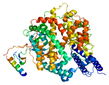
The ACE2 protein gene has 40 kb and 18 exons. Its protein has 805 amino acids with a molecular weight of approximately 120 kDa. Here, we have a view of the , going from the N-terminal to C-terminal region. Some other ways to represent the ACE2 protein include showing its , such as alpha helices and beta sheets, and mapping the polarity of its residues, differentiating and areas across its surface.
Besides its general aspects, the ACE2 protein can be divided into 4 portions: Peptidase Domain (Residues 19–615), Collectrin-like Domain (Residues 616–740), Transmembrane Domain (Residues 741–761) and Intracellular C-terminal Tail (Residues 762–805).
The is responsible for the enzymatic activity of ACE2. This domain can be divided in two subdomains: (residues 19–400) and (residues 401–615). Together, they form a substrate-binding cleft, where is located the , denominated HEXXH+E zinc-binding motif. Within this site, a zinc-ion is associated with the residues His374, His378, and Glu402, which are going to perform a nucleophilic attack on the peptide bond of the substrate, leading to its cleavage. within each subdomain, composed mainly of nonpolar residues provide tertiary structural stability, maintaining the correct spatial arrangement of catalytic residues. In the animation, we can observe a concentration of the hydrophobic residues towards the center of the molecule, while the polar ones are towards the outside part of the molecule.
An important region following the Peptidase Domain is the (CLD), also described structurally as a ferredoxin-like fold or neck domain, comprising residues approximately between 616 and 726. This domain connects the Peptidase Domain to the Transmembrane Domain and is composed of 4 beta sheets and 4 alpha-helices, arranged around a hydrophobic core. Its fold, characterized by a central beta-sheet flanked by alpha helices, plays a crucial role in stabilizing ACE2 dimers, which will be detailed later on this page.
The is composed of a single alpha helix, made of a majority of (in gray). This characteristic allows ACE2 to be anchored in the plasma membrane, due to its hydrophobic nature.
The final domain is the Intracellular C-terminal Tail but its function remains uncertain.
Physiological Function
is a transmembrane glycoprotein that has an extracellular catalytic domain. It is classified as a hydrolase, more specifically a carboxypeptidase-type peptidase, responsible for the cleavage of peptide bonds at the C-terminal site. [2]
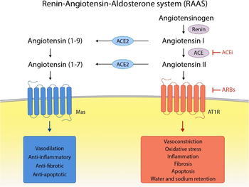
The ACE2 acts to cleave a single C-terminal residue of some substrates. One of those substrates is Angiotensin II (Ang II), which is degrated into Angiotensin-(1-7); to a lesser extent, Angiotensin I (Ang I) is also a substrate, turning in into Angiotensin-(1-9).
The Renin-Angiotensin-Aldosterone System - "RAAS" -, in which ACE2 acts, is an important signaling pathway responsible for vascular homeostasis.
In general, Ang II, generated by the action of ACE on Ang I, and this one by the action of Renin on Angiotensinogen, acts on AT1 and AT2 receptors. Its main action is vasoconstriction (by AT1 receptor), increasing blood pressure. On the other hand, Angiotensin (1-7), generated from Ang II by ACE2, has the opposite function, leading to a decline in blood pressure through vasodilation and release of Nitric Oxide (NO) through its action on the MAS oncogene receptor, antagonizing the actions of Ang II. [3]
 [4]
[4]
Pathological Relevance
The ACE2 protein became better known and widely publicized in 2020 due to its role in the infection by SARS-CoV-2, the virus responsible for the Covid-19 pandemic.
In this disease, ACE2 acts as a viral receptor, to which the Spike (S) viral protein binds, promoting the entry of the virus into the host cell. The specific region of the S protein responsible for binding to the ACE2 receptor is called receptor-binding domain (RBD), and is located in the S1 subunit of the Spike protein. [5]
Structure highlights[6]
ACE2-B0AT1 Complex
Association with B0AT1
The ACE2 protein can associate with the neutral amino acid transporter , also known as SLC6A19. In this context, ACE2 first forms a , where two ACE2 molecules interact side by side. Each ACE2 monomer then binds to one B0AT1 molecule. This results in a complex composed of two ACE2 and two B0AT1 molecules, which is commonly described as a . This association is essential for the transport of neutral amino acids in intestinal cells and the anchoring of ACE2 in the cell membrane, keeping the catalytic site facing the extracellular environment. Although ACE2 dimerization occurs independently of B0AT1, the transporter plays a stabilizing role by interacting with the Collectrin-like Domain.
This ACE2-B0AT1 complex is anchored to the cell plasma membrane, keeping the ACE2 protein in an extracellular environment. The intermembrane region has apolar characteristics, since it must interact with the hydrophobic tails of the phospholipids. Analyzing the of the complex, an apolar region is observed in the B0AT1 region, showing that it is in this region where the complex is in contact with the plasma membrane. The polar regions (CLD and PD domains of ACE2) are located in the external region, without contact with the apolar region of the phospholipids.
ACE2 Dimerization
ACE2 protein dimerization occurs independently of B0AT1 and involves both the Collectrin-like Domain (CLD) and regions from the Peptidase Domain (PD).
CLD Domain Interaction
In this region, there is an extensive network of polar interactions that stabilizes the ACE2 dimer. The most actively involved amino acid residues are between 636 and 658 and between 708 and 717, corresponding to the second and fourth helices of the CLD domain, respectively.
occur between the amino acids Arg652 and Tyr641 of the opposite ACE2 molecule, and between Arg710 and Tyr633. Additionally, help stabilize the dimer interface: Arg652 forms a hydrogen bond with Arg638, which further interacts with Gln653, linked to Asn636. Also, Arg710 forms a hydrogen bond with Asn639, and Arg716 connects with Ser709 and Asp713.
Domain PD Interation
In addition to the CLD, the Peptidase Domain (PD) also contributes to ACE2 dimerization. The amino acids Cys133, Asn134, Asp136, Asn137, Gln139, Glu140, and Cys141 form a region within the PD. The two cysteines (Cys133 and Cys141) establish a disulfide bond, which stabilizes the loop conformation, supported by additional intraloop polar interactions. Furthermore, Gln139 forms a polar interaction with Gln175 from the other ACE2 monomer.
SARS-CoV-2 Binding
ACE2 to the Spike (S) glycoprotein of the SARS-CoV-2 virus, promoting its internalization into the cell. More specifically, binding occurs between the PD subunit of ACE2 and the receptor binding domain (RBD) of the S1 subunit of the S protein. Each PD binds to an RBD by polar bonds, with one ACE2 dimer accommodating two S protein trimers.


