Print3D help
From Proteopedia
Proteopedia and 3D Printing
Physical molecular models, by engaging both visual and tactile senses, provide an effective means of deepening the understanding of complex concepts in molecular biology and biochemistry. They allow students to perceive the three-dimensional organization of macromolecules, thereby enhancing comprehension of the relationship between structure and function [1] [2]. Numerous studies have demonstrated that the use of tangible models in science education improves learning outcomes, fosters conceptual reasoning, and increases student engagement [3] [4] [5]. The rapid development of 3D-printing technologies has made it possible to create customized and affordable molecular models [6] [7] [8].
Affordable desktop 3D printers can now be used to fabricate physical models from virtually any structure or scene on Proteopedia. This page provides an introduction to 3D printing, a step-by-step guide to generating printable files directly from Proteopedia, and several tested printing profiles for common printers (e.g., Prusa Research, Bambu Lab, Creality, or Elegoo).
Let’s 3D-print the whole Proteopedia!
«3D Printing 101»: an introduction to 3D printing
3D printing, also known as additive manufacturing, builds physical objects layer by layer from digital designs. For more technical details please head for the excellent introductory article on All3DP. In structural biology education, 3D printing provides an affordable and accessible way to transform atomic coordinate data (e.g., from PDB, MOL or SDF files) into tangible molecular models that can be observed, handled, and discussed in class or laboratory settings.
Printer Types: FDM vs. SLA/DLP
The two most accessible 3D-printing technologies that can be used to create molecular models are:
- FDM (Fused Deposition Modeling)
This method extrudes a heated thermoplastic filament (such as PLA, PETG or TPU) through a fine nozzle, depositing successive layers that form the model. FDM printers have the advantages of low cost, easy maintenance, wide availability, and being capable of printing large and colorful molecular models. This technology has a few limitations, such as: visible layer lines, lower surface resolution, and less suited for very small or intricate molecular details.
- SLA/DLP (Resin Printing)
This technique uses liquid photopolymer resin, cured layer by layer with ultraviolet light. SLA/DLP printers have the advantages of a very high resolution, smooth surfaces and are ideal for compact and detailed models (e.g., small proteins rendered as surface, ligands, or symmetric assemblies). This technology has its own limitations, such as higher material cost, post-processing requirements (washing and UV-curing), and the need for careful handling of resin.
When choosing between FDM and SLA printers, model size and resolution are the key criteria: FDM is preferred for large, educational display models; SLA/DLP is optimal for small, precise structures.
While FDM and SLA/DLP are the most widely used technologies in educational and research contexts due to their accessibility, other additive manufacturing methods also exist —such as Selective Laser Sintering (SLS), PolyJet, and Binder Jetting— each employing different materials and curing mechanisms. These industrial-grade techniques can produce highly detailed or multi-material parts but are generally more expensive and less accessible for classroom use. For an overview of major 3D-printing technologies, see All3DP: Complete Overview of the Types of 3D Printers
Single- vs. Multi-Material Printers
Most entry-level desktop 3D printers use a single filament or resin, producing models in one color or material. However, multi-material systems (such as Bambu Lab AMS and its variants or Prusa MMU3) can simultaneously print multiple filaments, enabling color-coded domains, subunits, or ligands directly in one print. For complex color information, users can also apply post-print color coding (painting or labeling) or print separate parts and assemble them manually.
| A large FDM-printed model | A multi-material FDM-printed model | Small resin-printed models |
|---|---|---|
 GABAA receptor, 6x3x, scale at the bottom in cm |  Bacterial 70S ribosome, 4v5d, scale at the bottom in cm | |
| Trace Model of the GABAA receptor printed on a single material FDM-Printer (Prusa MK3S+). Each of the 5 subunits was printed separately and can be assembled. Printable files available on Printables.com | Surface model of the Bacterial 70S ribosome printed on a multi-material capable printer (BambuLab X1C AMS). Blue and violet are ribosomal proteins, orange and yellow are rRNAs, and black is tRNA. Printable files available on Printables.com | Balls and sticks and cartoon models of DNA (right) and insulin (left) |
File Formats Used in 3D Printing
Proteopedia supports two export formats for generating printable molecular models:
- .STL (Stereolithography): the most common and universally compatible format; encodes only surface geometry, without color or texture information.
- .OBJ (Wavefront Object) with an associated .MTL (Material Template Library) files: includes both geometry and optional color/texture data; useful for color prints.
Another widely adopted file format used in 3D printing is the .3MF (3D Manufacturing Format): a modern open standard that stores geometry, color, and multi-material metadata in a single file—recommended for advanced printers. For a more extensive pros and cons of different 3D printing file formats please see The Main 3D Printing File Formats on all3dp.
STL is ideal for single-color models and maximum compatibility, while OBJ/MTL is recommended for multi-material workflows. 3MF files will be used in this tutorial to save the model and printing settings.
Support in Proteopedia for these formats is based on the export capabilities of Jmol/JSmol; for details, see STL and OBJ
Overview of the 3D printing process
3D printing enables the transformation of atomic-level structural data into tangible macromolecular models that can be explored through both sight and touch. The process generally follows several main steps, each requiring specific types of software and hardware.
Preparing the structure and generating a printable File
- The first stage involves creating a printable three-dimensional representation of the molecule. Some molecular-graphics programs such as PyMOL, UCSF ChimeraX, [1], or Jmol can be used not only to visualize a structure (from the Protein Data Bank or other sources), but also to export the resulting geometry in a .STL or .OBJ format. However, the default visualization styles of these programs are not directly suitable for 3D printing. Therefore, when preparing a model for fabrication, it is important to:
- Select an appropriate molecular representation (e.g., surface, cartoon, sticks, spheres) depending on the pedagogical or structural feature you wish to highlight.
- Add struts or connectors to increase the mechanical stability of the model, especially for large complexes or long helices.
- Remove specific struts or pseudobonds when individual components — ligands, subunits, or cofactors — need to remain detachable.
- Adjust thickness of atoms, bonds, helices, and sheets to ensure adequate rigidity and printability (features thinner than the printer’s nozzle diameter or resin resolution may break).
- Apply meaningful coloring when preparing multi-material prints, for instance, to distinguish domains, chains, ligands or atoms.
Proteopedia integrates a dedicated Print3D tool within each molecular scene that automates the process and enables users to effortlessly generate 3D-printable molecular models - more on that in the corresponding section Generating printable files on Proteopedia within this page. For advanced users wanting to do this manually, we highly recommend two guides: [6] and [7].
Slicing the Model
Before printing, the STL file must be processed by a slicer — specialized software that converts 3D geometry into a series of horizontal layers. The slicer also generates G-code, the set of instructions that control the printer. These commands are printer-specific and include parameters such as extrusion speed, layer thickness, temperature, and movement. Together, they are usually stored as a printer profile.
During the slicing of a molecular model for an FDM printer, users can adjust:
- number of perimeters, which affects mechanical strength and print time;
- layer height, which determines resolution and print duration;
- infill density, which determines internal strength and affects print time;
- supports, temporary structures that hold overhanging regions;
- print orientation, which influences surface quality, mechanical strength, and the amount of supports required.
- All these parameters make up a print settings profile or process profile. In this step, users must also select the material-specific parameters, or apply a filament profile. More details on these profiles are provided later on this page.
During the slicing of a molecular model for an SLA/DLP printer, users can adjust:
- layer height, which determines vertical resolution and print time; thinner layers produce smoother surfaces but significantly increase printing duration.
- exposure time, which controls how long each layer is cured by UV light; optimal exposure ensures proper adhesion between layers without over-curing fine details.
- bottom-layer exposure and count, which influence model adhesion to the build platform — longer exposures and additional base layers improve stability during printing.
- supports, which anchor the model to the build plate and prevent deformation during peeling; proper placement reduces surface marks and print failures.
- model orientation, which affects surface quality, support contact area, and resin drainage; angling the model (typically 30–45°) minimizes suction forces and improves print reliability.
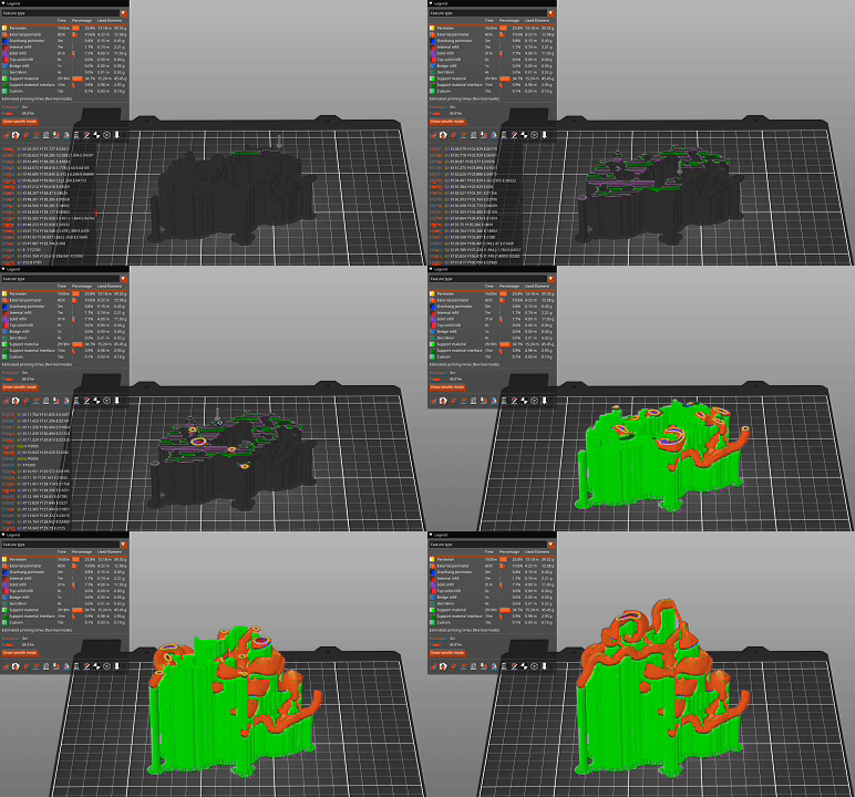
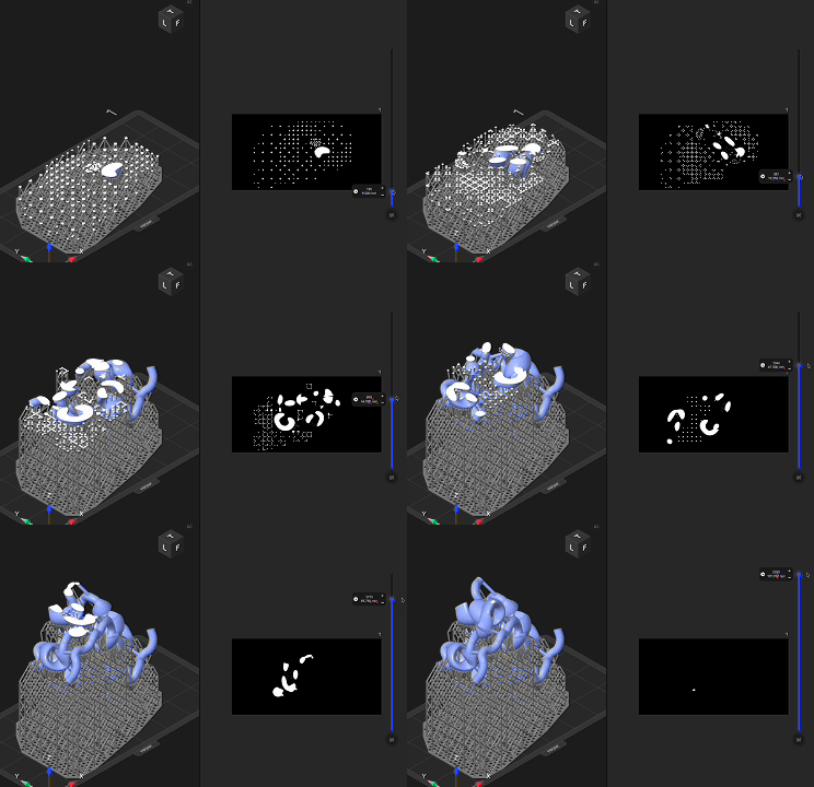
| An insulin model sliced for FDM printing | An insulin model sliced for resin printing |
|---|---|
 Cartoon model of insulin, 4ins |  Cartoon model of insulin, 4ins |
| Cartoon model of insulin sliced in PrusaSlicer and ready to be printed on FDM printers. The arrow shows the trajectory of the print head on a single slice or layer. Orange is the model, green are the supports. | Cartoon model of insulin sliced in Elegoo SatelLite and ready to be printed on FDM printer.On the left, with blue is the model, with gray are the supports. On the right is the image on the UV screen. |
3D Printing the Model
- The prepared G-code is then transferred to the 3D printer, either via an SD card, USB flash drive, or Wi-Fi connection. During printing, close observation of the first few layers is essential to ensure good adhesion to the build plate and a successful print. Total printing time depends on model size, selected layer height, and printer speed. Small peptide models may print in less than an hour, whereas large multimeric assemblies can require several hours or days. Multi-color or multi-material prints generally take longer, while SLA/DLP printers are generally faster due to their simultaneous layer exposure.
Post-Processing
- After printing, the model undergoes post-processing, which varies depending on the printing technology used:
- For FDM prints, supports are removed mechanically, and surfaces can be smoothed by light sanding or polishing.
- For resin prints, supports are removed mechanically, parts are washed in isopropanol to remove uncured resin and then exposed to UV light for final curing.
- Models printed in multiple parts can be assembled using cyanoacrylate or epoxy adhesive. Optional finishing steps (painting, labeling) can improve contrast between subunits or highlight functional regions.
Generating printable files on Proteopedia
Proteopedia integrates a dedicated Print3D tool within each molecular scene, enabling logged-in users to effortlessly generate 3D-printable molecular models directly from its pages. The tool is powered by 3DP-Jmol, a script that analyzes the structural data and automatically scales the model’s geometry to match the printable volume of a medium-size desktop 3D printer (e.g., Ender-3, 210×210×210 mm). During this process, the tool evaluates the molecular bounding box, atom radii, and bond lengths, then proportionally adjusts all dimensions to achieve a realistic and mechanically robust print scale — preserving scientific accuracy while ensuring manufacturability. For advanced users, 3DP-Jmol may also be used in conjunction with the Jmol program, providing full access to its scripting capabilities and advanced export features.
Step-by-step guide for automatic generation of printable files on Proteopedia
Open a structure and access the Print3D tool
Log in with your Proteopedia Username and password. Navigate to any Proteopedia page (for example, Introduction_to_protein_structure). The corresponding 3D structure will load in the viewer on the right.
At the bottom of the structure display window, locate and click the Print3D button. The tool will launch and begin analyzing the current molecular structure. This process may take some time to complete, depending on the size of the structure. Please wait until the message Analyzing structure... disappears and the next page is displayed — this marks the start of the two-step printing workflow.
Select a molecular representation and a printing scale
In this new page, some user input is required. Each option includes a short explanation; the key steps are outlined below, but feel free to explore and adopt the settings that best fit your use case.
(1) Choose a molecular representation from the Scheme list, based on your goal:
- Backbone, Trace, or Ribbon — to emphasize secondary structure (effective for teaching protein folds).
- Surface — to highlight molecular topology and binding sites.
- Ball & Stick — atom-level detail for ligands or small molecules; usually impractical for large proteins or complexes.
- Proteopedia Scene — attempts to render the current page’s scene directly; may not work for all scenes. This is not fully implemented and only available for a few users.
Note: Some representations may be unavailable for a given structure because they are not printable in a reliable way.
(2) In the Printer section, select whether you want a single-color or a multi-color print.
(3) In the Printer scale section, set the desired size of the printed model. The tool constrains safe sizes based on the chosen representation and displays the physical dimensions (mm). Important! Please note the printer scale value in % as it is going to be required downstream in the next section of this workflow, Printing files from Proteopedia.
(4) When ready, click the green Make printer files button. Please wait patiently until the Generating files for 3D printing of... message disappears and the final step is displayed.
Download the .STL or .OBJ files
In this final page, a 3D preview of the generated model is displayed. You can use the mouse to rotate, zoom, and inspect the model from different angles. If the result meets your needs, click the green (A) Download files button and save the compressed .zip archive containing the .STL or .OBJ files on your computer. If your model needs further improvements, hit the green (B) Same structure button to return to the previous screen and adjust your settings.
Printing files from Proteopedia
Unzipping the compressed .zip archive will produce the exported .STL or .OBJ file, along with an optional .MTL material file.
Using 3D printing services
If you do not have a 3D printer, you can still obtain physical models by using locally available 3D-printing services. Just search on Google Maps with the term 3D printing in your area. Here is short list of well-known 3D Printing service providers that may also ship worldwide:
- Shapeways — global service offering a wide range of materials and colors. Educational discounts are available through their academic program.
- Sculpteo — Europe-based provider offering fast turnaround and many printing technologies, including resin and polymer options.
- Xometry — large international network providing access to industrial-grade printing methods such as FDM, SLA, SLS, and DMLS.
- Craftcloud — an aggregator that compares multiple providers and finds the best price and lead time for your uploaded model.
When contacting a printing service, provide the exported files together with the printer scale value in % saved in the previous step (Select a molecular representation and a printing scale). It is also advisable to share the link to this Proteopedia page so that the operator understands the peculiarities of printing molecular models.
Using a desktop 3D printer
Before importing the .STL or .OBJ file into your slicer software, it is recommended to inspect and, if necessary, repair the model to ensure printability. Common issues include non-manifold geometry, intersecting faces, and missing surfaces. The example below demonstrates how to use Blender together with its 3D Print Toolbox extension to improve the printability of the .STL and .OBJ files generated with the Print3D tool in Proteopedia. Important note: The repair process is largely automated but may introduce artifacts. Carefully inspect the corrected model for any missing or distorted sections, atoms, or bonds, and determine whether these omissions affect the intended purpose of the printed model.
| How to fix the molecular models generated by Proteopedia for single color or resin printing | How to fix the molecular models generated by Proteopedia for multi-color FDM printing |
|---|---|
Once the repaired .STL or .OBJ file is ready, it can be imported into your slicer software. In this step, you will prepare the file for printing by applying appropriate printer, filament, and print-settings profiles.
- Apply the printer scale value (%) saved in the previous step ( Select a molecular representation and a printing scale). This step ensures that the model fits your print bed and maintains structural integrity during printing. The location of the scale (%) input field varies depending on the slicer software you are using.
- Orient the model on the print bed so that its largest, flattest surface rests on the bed. This minimizes the need for supports and improves adhesion. If your slicer includes an Auto Orient or Auto Arrange function, use it to optimize placement. (Hint: Bambu Studio and Orca Slicer include this feature, Ultimaker Cura has a Auto-Orientation plugin available, while PrusaSlicer currently does not.)
- Select the printer by applying a printer profile that matches your device.
- Choose the material by applying a filament profile corresponding to the filament type (e.g., PLA, PETG, TPU) and manufacturer you intend to use.
- Adjust the printing parameters by selecting a print settings profile or process profile that defines layer height, infill, speed, and support options.
In most cases, the default printer profile and filament profile provided by the slicer are sufficient. However, molecular models often include overhangs, thin connections, and complex internal geometries, which require additional supports and reinforced perimeters to ensure structural stability. For this reason, the default print settings profile or process profile may not yield optimal results.
Validated printing profiles for the Print3D tool are available for several printer models. These profiles provide optimized settings that produce sturdy models, reduce material consumption, and shorten print time, ensuring consistent and reliable results. Three distinct profiles are supplied, corresponding to major molecular representations: balls and sticks, surface, and cartoon. The cartoon profile is also suitable for backbone, trace, and ribbon representations. For some printers, each profile is supplied in two variants, one using standard supports and one using tree or organic supports. In the case of multi-material printers, profiles for printing with soluble support and soluble interface are also provided.
| Printer brand and model | Profiles provided as / Slicer | Printer Type, nozle size and multi-material support | GitHub with profiles | How to import profiles into Slicer |
|---|---|---|---|---|
| Original Prusa Core One | a single profile per .INI file / PrusaSlicer | FDM / 0.4 mm / yes, 5 colors, if MMU3 installed | CoreOne | Import and export custom profiles in PrusaSlicer |
| BambuLab A1Mini | a single Process preset .zip file with all presets / Bambus Studio | FDM / 0.4 mm / yes, 4 colors if AMS Lite is installed | AiMini | Import and export custom profiles in Bambus Studio |
| BambuLab X1 Carbon | a single Process preset .zip file with all presets / Bambus Studio | FDM / 0.4 mm / yes, up to 16 colors if multiple AMS are installed | X1Carbon | Import and export custom profiles in Bambus Studio |
| Original Prusa MK3S+ | a single profile per .INI file / PrusaSlicer | FDM / 0.4 mm / no | MK3S+ | Import and export custom profiles in PrusaSlicer |
| Creality Ender 5 Pro | a single profile per .INI file / PrusaSlicer | FDM / 0.4 mm / no | Ender5Pro | Import and export custom profiles in PrusaSlicer |
| Creality Ender 3 v2 with a ENDERIDEX kit | a single profile per .INI file / PrusaSlicer | FDM / 0.4 mm / yes, IDEX, two filaments | Ender3IDEX | Import and export custom profiles in PrusaSlicer |
| Kingroon KPS3 Pro | a single profile per .INI file / PrusaSlicer | FDM / 0.4 mm / no | Ender3IDEX | Import and export custom profiles in PrusaSlicer |
| Elegoo Mars 5 Ultra | no profile needed, just be sure to add supports / ELEGOO SatelLite | resin / - / no | no profile needed, default settings worked, just be sure to add supports. EVO supports worked well enough. Post-processing steps: 1)washing: at least 10 min. 2) support removal 3)curring: at least 10 min on a Elegoo Mercury Plus V3.0 |
Notes:
- These profiles were developed and validated with the support of EDUMOL3D, Project PN-IV-P7-7.1-PED-2024-0343 financed by the Ministry of Research, Innovation and Digitization, UEFISCDI, Romania.
- The profiles are a work in progress. Please check GitHub for updates. If you have suggestions for improvements, please drop a message at marius.mihasan(@)uaic.ro
Note on multi-color printing
To our knowledge, only Bambu Studio and Orca Slicer can automatically import color information from the .MTL file associated with the printable .OBJ generated by the Proteopedia Print3D tool. At present, PrusaSlicer does not support importing .MTL files directly.
A practical workaround for PrusaSlicer users is as follows:
- Open the .OBJ file in Bambu Studio.
- Verify or adjust the color mapping as needed.
- Export the model as a generic .3MF file using the menu command: File → Export → Export Generic 3MF...
- Open the exported .3MF file in PrusaSlicer. The color data will now be available under the Multimaterial Painting tool.
This workflow preserves the original coloring assigned in Proteopedia and allows successful multi-color or multi-material printing on any capable 3D printer compatible with PrusaSlicer.
Other relevant resources
Print3D Models gallery - examples of 3D printed molecular models from Proteopedia users.
NIH 3D Print Exchange - an open, community-driven portal to download, share, and create bioscientific and medical 3D models, including for 3D printing.
3DP-Jmol - the Jmol/JSmol behind the Print3D tool. Automatically generates 3D printable molecular models from structural data and can be easily integrated into web pages.
3D Printing on PDB101 - a quick guide on 3D printing molecular models and a small curated 3D models for education
If you print a model from Proteopedia, please share photos and feedback — let’s 3D print the whole Proteopedia!
References
- ↑ Howell ME, Booth CS, Sikich SM, Helikar T, van Dijk K, Roston RL, Couch BA. Interactive learning modules with 3D printed models improve student understanding of protein structure-function relationships. Biochem Mol Biol Educ. 2020 Jul;48(4):356-368. PMID:32590880 doi:10.1002/bmb.21362
- ↑ Răzvan-Ştefan B, Laura Nicoleta P, Mihășan M. Impact of 3D-printed molecular models on student understanding of macromolecular structures: a compensatory research study. Biochem Mol Biol Educ. 2025 Jul-Aug;53(4):358-369. PMID:40214166 doi:10.1002/bmb.21902
- ↑ Smith DP. Active learning in the lecture theatre using 3D printed objects. F1000Res. 2016 Jan 13;5:61. PMID:27366318 doi:10.12688/f1000research.7632.2
- ↑ Srivastava A. Building mental models by dissecting physical models. Biochem Mol Biol Educ. 2016 Jan-Feb;44(1):7-11. PMID:26712513 doi:10.1002/bmb.20921
- ↑ Larsson, C., Tibell, L.A.E. Challenging Students’ Intuitions—the Influence of a Tangible Model of Virus Assembly on Students’ Conceptual Reasoning About the Process of Self-Assembly. Res Sci Educ 45, 663–690 (2015). DOI: 110.1007/s11165-014-9446-6
- ↑ 6.0 6.1 Da Veiga Beltrame E, Tyrwhitt-Drake J, Roy I, Shalaby R, Suckale J, Pomeranz Krummel D. 3D Printing of Biomolecular Models for Research and Pedagogy. J Vis Exp. 2017 Mar 13;(121):55427. PMID:28362403 doi:10.3791/55427
- ↑ 7.0 7.1 Mihasan M. A beginner's guideline for low-cost 3D printing of macromolecules usable for teaching and demonstration. Biochem Mol Biol Educ. 2021 Jul;49(4):521-528. PMID:33755300 doi:10.1002/bmb.21493
- ↑ Segarra VA, Chi RJ. Combining 3D-Printed Models and Open Source Molecular Modeling of p53 To Engage Students with Concepts in Cell Biology. J Microbiol Biol Educ. 2020 Dec 21;21(3):21.3.72. PMID:33384761 doi:10.1128/jmbe.v21i3.2161


