User:Saad Siddiqui/Sandbox 1
From Proteopedia
Contents |
Rhodopsin
Introduction
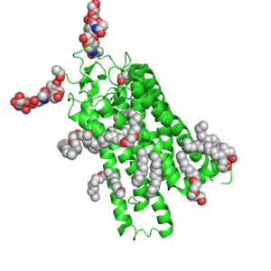
Rhodopsin is a G-protein coupled receptor that sits in the membrane of the outer segment of retinal rod cells in the eye. The outer segment in mammals is made of thousands of disks, which are made up almost entirely of rhodopsin. Each rhodopsin molecule spans the disk seven times in a typical transmembrane helical structure. A light absorbing molecule called retinal is attached to seventh helix. Retinal absorbs a photon of light and changes its structure, which induces a change in the rhodopsin protein. This triggers a series of events in the transduction of light to a signal in the brain, and is the basis for vision.
Background
The rich history of rhodopsin predates its biochemical discovery. To understand the importance of rhodopsin it is imperative that one consider the process which it inaugurates. Vision has been the subject of intense philosophical and scientific inquiry since the Greek era.[1]Early Greek philosophers proposed several optical theories regarding the nature of light transmission in the eye. The great physician Galen drew detailed diagrams of the eye and proposed a theory connecting the eye and the brain.[2] In the middle ages, Alhazen conducted experiments to test the validity of the various theories.[3] In 1704, Newton published his Opticks, revealing new insights about the physical nature of light.
The biological history of rhodopsin is intertwined with the knowledge of night blindness and the discovery of vitamin A.[4] Physicians in the Greek era were aware that night blindness was a physiological condition that could be cured with proper nutrition. Two thousand years later, in 1742, van Leewenhoek described rods and cones within the eye.[4] Muller noted that the rods were pigmented, but he thought this was due to hemoglobin. In 1851, Franz Boll experimented on the rod cells of frogs. He noticed that when he removed the retina, the pigment in the rods faded away within a minute. He established that this was not due to death but because light was striking the cells. Boll died before he could isolate the substance. It was Willy Kuhne who took up the study of rod cells in 1877.[4] He was the first to name the pigmented substance rhodopsin, because he observed it was purple.[5] He postulated that rhodopsin was a protein, and that there were actually two substances involved in the reaction with light.[5] Kuhne was the first to extract rhodopsin and work out several important steps in the photochemical pathway of vision transduction.[6]
It was not until the 1930’s that the relationship between rhodopsin, vitamin A, and night blindness was pieced together. George Wald recognized that the absorption spectrum of retinas was reminiscent of vitamin A. He read previous literature which related vitamin A deficiency with night blindness and recognized that it was involved in the process of vision transduction. With the help of organic chemists, Wald and others were able to show that the light absorbing compound was 11-cis-retinal, and this combined with one opsin to make rhodopsin.[5] His lab showed that 11-cis-retinal was converted to all-trans-retinal and they theorized that this triggered a cascade of reactions to transduce the signal from a photon.[5]
With the advance of x-ray crystallization and sequence comparison, the structure of rhodopsin was carefully elucidated throughout the 1990’s. Sequence comparisons had established that it was a model GPCR protein, but the location of the helices was unknown.[7] From projection maps, low resolution 3-D structures were obtained by Unger and Schertler.[7] In the past decade, very high resolution images of rhodopsin have been crystallized, allowing biochemists to fully appreciate the its role in vision.[8]
Structure
|
Rhodopsin shares its structure with a family of proteins known as G-protein coupled receptors.[9] GPCR structure is one of the most highly conserved through evolution, and over 900 genes in the human genome encode GPCR’s.[8] The of GPCR’s is that of seven transmembrane domains with three intracellular, and three extracellular loops on either side of the membrane.[9] The structure of rhodopsin has been found in the phototaxis machinery of bacteria.[10] The first “eyespots” in bacteria allowed the detection of light, and movement in that direction. It was found in 1982 that the phototaxis receptor in the prokaryote Halobacterium salinarum, is a seven-pass transmembrane protein which uses a retinylidene chromophore.[10] It is unclear whether this receptor and the animal receptor share a common ancestor or not, but the similarity speaks to the utility of the rhodopsin structure in relation to signal transduction.
Mammalian rhodopsin is a GPCR with a weight of 40,000.[11] Approximately 50% of the peptides in the rhodopsin sequence are hydrophobic, which indicates the transmembrane architecture.[12] The seventh helix is linked to 11-cis-retinal at the Lys-296 residue by a protonated Schiff base.[8] The , which serves as the chromophore, is embedded within the middle of the bilayer.[11] The second extracellular loop effectively where retinal binds.[8] A disulfide bond between the Cys-187 of the loop an Cys-110 of helix III forms this “seal”.[8] Furthermore, the terminal residues of extracellular loop II (Met183 and Gln184) have side chains which extend into the center of the protein to form a hydrophobic pocket.[10] A beta sheet B4 located below the retinal molecule forms the binding pocket.[10] Interestingly, the extracellular face of rhodopsin is much more compact and organized. Several interactions account for this. Strand S4 connects the Ser14-Asn15 in the terminal region with Pro23. Strand 5 covers the space between helices 1 and 2.[10] Helices I, IV, VI, and VII are bent at proline residues, while helix V is straight, in the middle of the protein. Helix II is bent such that Gly90 is close to the base Glu113.
The retinal chromophore has been identified as 6s-cis, 11-cis, 12s-trans, antiC=N. This molecule is bound to the protein at the Lys296 residue with a protonated Schiff base. The orientation of the Lys296 side chain is bounded by the hydrophobic side chains of Met44 and Leu47 and further stabilized phenyl rings which interact with the helices.[10]
The cytoplasmic region contains a loop (loop II) which marks a border from the transmembrane region.[10] Loop III has been shown to interact , the G-protein that associates with rhodopsin.[10]
Function
The function of proteins in the GPCR family has been conserved along with the general structure. The mechanism of signal transduction begins when the GPCR receives a signal from the outside. The receptor stimulates a guanosine nucleotide-binding protein (G protein), which in turn activates an effector enzyme. The effector enzyme induces a second messenger which amplifies the signal.
Rhodopsin’s primary role in mammalian cells is phototransduction.[13] Simply put, rhodopsin is a molecular switch which is turned “on” when it receives a visual signal in a photon of light. The 11-cis-retinal that is attached to the opsin isomerizes to all-trans-retinal. The structure of the chromophore-protein binding induces changes in the opsin as well, turning it “on” in an active state known as metarhodopsin.[13] The activated rhodopsin can then binds the G-protein transducin in order to activate it.[13]
When light strikes the eye, the individual photons are taken up by the rod cells and they induce the cis-to-trans isomerization of retinal. Though this reaction is complete within femtoseconds, several intermediate states of rhodopsin have been observed. Each intermediate state has been identified by its absorption spectrum.[12] During these intermediate states, rhodopsin is able to interact with many different proteins to do its work. These intermediate steps are necessary functionally because they are on the path to the active molecule which can bind transducin.[12] One of the first steps is the deprotonation of the Schiff base, and the protonation of Glu113 (which accepts the proton).[14] This step disrupts a salt bridge which normally stabilizes the ground state rhodopsin.[14] Other factors are important for the activation of rhodopsin, such as protonation of Glu134.
Once rhodopsin is turned on, it can activate transducing constitutively until it is turned off.[15] Rhodopsin kinase phosphorylates rhodopsin, allowing visual arrestin to bind as well. This effectively shuts off the activation of transducin. To regenerate rhodopsin, the all-trans-retinal is lost, followed by arrestin, and the rhodopsin is dephosphorylated. 11-cis-retinal is regenerated and the switch is ready once more.
Role in Disease
Mutations in rhodopsin cause various types of retinal diseases.[14] The first identified mutation was a Pro23His substitution which was found to be associated with retinal degeneration.[14] Since then, hundreds of mutations have been found to cause various forms of the disease. Protein misfolding can occur, leading to loss of function. When an abnormal disulfide bond is formed between Cys185 and Cys187, for example, the protein becomes misfolded.[14]
Another structural mutation disrupts crucial function causing CSNB.[14] When Gly90 and Thr94 are mutated, they disrupt the salt bridge which normally forms between the protonated Schiff base and Glu113. This salt bridge normally stabilizes the ground state rhodopsin, and disruption causes a malfunction in the protein.
Many different types of mutations have been identified and there have been proposals for classifications based on the mechanism by which the mutation inhibits function.[16]At least 7 classes of mutants have been proposed, based on if they affect folding, action of the opsin, or isomerization of retinal.[16]
It is crucial to understand disease induction due to rhodopsin malfunction because rhodopsin is one of the most studied GPCR’s in mammals. The results of research on retinal disease and rhodopsin function will prove fruitful in countless other areas.
PDB Entry
3C9L is an alternative model with sequence from Bos taurus. Two relevant features are the March 2002 RCSB PDB Molecule of the Month on Bacteriorhodopsin and the October 2004 Molecule of the Month on G Proteins, both by David S. Goodsell. Full crystallographic information is available from OCA.
References
- ↑ [1]Wade, N.J. A Natural History of Vision. MIT Press: Cambridge, 1999. 1-20.
- ↑ [2]Cherniss, H. Galen and Posidonius' Theory of Vision. Am. J. Philology. 1933, 54(2), 154-161.
- ↑ [3]Lindberg, D.C. Alhazen's Theory of Vision and its Reception in the West. Isis. 1967, 58(3), 321-341.
- ↑ 4.0 4.1 4.2 [4]Wolf, G. The discovery of the visual function of vitamin A. J. Nutr. 2001, 131(6), 1647-1650.
- ↑ 5.0 5.1 5.2 5.3 [5]Hubbard, R.; Wald, E. George Wald memorial talk. 1999. Rhodopsins and phototransduction. Wiley, Chichester (Novartis Foundation Symposium) 5-20.
- ↑ [6]Palczewski, K. G Protein-Coupled Receptor Rhodopsin. Annu. Rev. Biochem. 2006, 75, 743-767.
- ↑ 7.0 7.1 [7]Schertler, G.F. Structure of Rhodopsin. 1999. Rhodopsins and phototransduction. Wiley, Chichester (Novartis Foundation Symposium) 54-69.
- ↑ 8.0 8.1 8.2 8.3 8.4 [8]Ridge, K.D.; Palxzewski, K. Visual Rhodopsin Sees the Light: Structure and Mechanism of G Protein Signaling. J. Biol. Chem. 2007, 282, 9297-9301.
- ↑ 9.0 9.1 [9]Rompler, H.; Staubert, C.; Thor, D.; Schulz, A.; Hofreiter, M.; Schoneberg, T. G protein-coupled time travel: evolutionary aspects of GPCR research. Mol. Interv. 2007, 7(1), 17-25.
- ↑ 10.0 10.1 10.2 10.3 10.4 10.5 10.6 10.7 [12]Palczewski, K.; Kumasaka, T.; Hori, T.; Behnke, C.A.; Motoshima, H.; Fox, B.A.; Le Trong, I; Teller, D.C.; Okada, T.; Stenkamp, R.E.; Yamamoto, M.; Miyano, M. Crystal structure of rhodopsin: A G protein-coupled receptor.Science. 2000, 289(5480), 739-745.
- ↑ 11.0 11.1 [10]Nelson, D.L.; Cox, M.M. Principles of Biochemistry. W.H Freeman: New York, 2008. 461-469.
- ↑ 12.0 12.1 12.2 [11]Stavenga, D.G.; DeGrip, W.J.; Pugh, E.N. Molecular Mechanisms in Vision Transduction. Elsevier: Amsterdam, 2000. 1-18.
- ↑ 13.0 13.1 13.2 [13]Hargrave, P.A. Rhodopsin structure, function, and topography the Friedenwald lecture. Invest. Opthalmol. Vis. Sci. 2001, 42(1), 3-9.
- ↑ 14.0 14.1 14.2 14.3 14.4 14.5 [15]Garriga, P; Manyosa, J. The eye photoreceptor protein rhodopsin. Structural implications for retinal disease. FEBS Lett. 2002, 528, 17–22.
- ↑ [14]Fliesler, S.J.; Kisselev, O.G. Signal Transduction in the Retina. CRC Press:Boca Raton, Fl, 2008. 35-39.
- ↑ 16.0 16.1 [16]Rakoczy, E.P.; Kiel, C.; MeKeone, R.; Stricher, F.; Serrano, L.; Analysis of Disease-Linked Rhodopsin Mutations Based on Structure, Function, and Protein Stability Calculations. J. Mol. Biol. 2011, 405(2), 584-606.

