User:Tiana Yom/sandbox1
From Proteopedia
Cyan Fluorescent Protein
The protein responsible for the bioluminescence also known as green fluorescent protein (GFP) was discovered by scientists in Aequorea victoria. Since then, GFP has revolutionized the field of science and has allowed scientists tag cells in order to view them in vivo. The chromophore, which contains the Ser65-Tyr66-Gly67, is stabilized by the beta barrel that surrounds the structure. GFP contains only one tryptophan amino acid (Trp66) in the whole protein. Simple modifications to this sequence can allow GFP to undergo several different color changes. When Tyr66, in the same position of the chromophore, changes into another tryptophan amino acid the color of GFP changes to Cyan Fluorescent Protein (CFP). This change allows for the protein to fluoresce blue. The Tyr66 in the GFP molecule plays a primary role in stabilizing the green fluorescent. This is due to the aromatic rings that the amino acid contains. Likewise, Tryptophan contains aromatic rings. In the PDB model 1oxe of CPF, we highlighted the specific amino acid that is pertinent in color change from green to blue. The amino acid, tryptophan, is colored dark blue in the rasmol model. Along with tryptophan, we used a light blue color to display the nitrogen in the tryptophan indole ring. A minute change in amino acid Tyr66 allowed for the fluorescent protein to go from green to blue. The importance of these GFP derivatives is that we are able to trace various types of cells at the same time in vitro.
Replace the PDB id (use lowercase!) after the STRUCTURE_ and after PDB= to load and display another structure.
| |||||||||
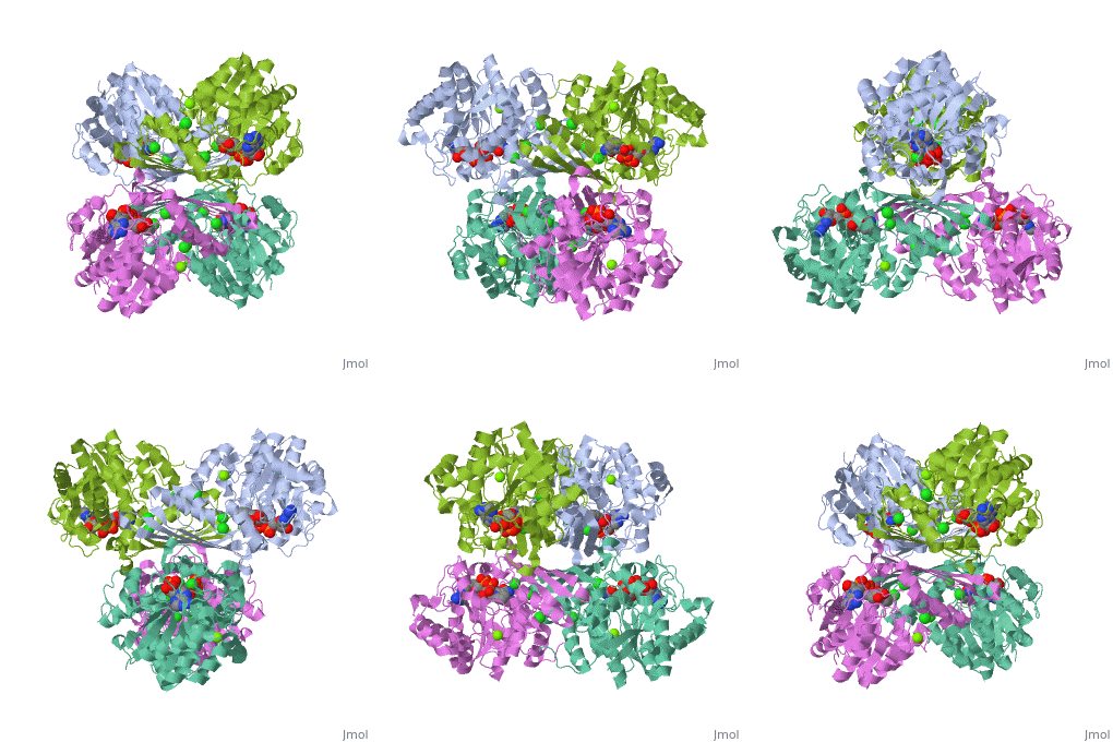
| 3cin, resolution 1.70Å () | |||||||||
|---|---|---|---|---|---|---|---|---|---|
| Ligands: | , , | ||||||||
| Gene: | TM1419, TM_1419 (Thermotoga maritima MSB8) | ||||||||
| Activity: | Inositol-3-phosphate synthase, with EC number 5.5.1.4 | ||||||||
| |||||||||
| |||||||||
| Resources: | FirstGlance, OCA, RCSB, PDBsum, TOPSAN | ||||||||
| Coordinates: | save as pdb, mmCIF, xml | ||||||||


