We apologize for Proteopedia being slow to respond. For the past two years, a new implementation of Proteopedia has been being built. Soon, it will replace this 18-year old system. All existing content will be moved to the new system at a date that will be announced here.
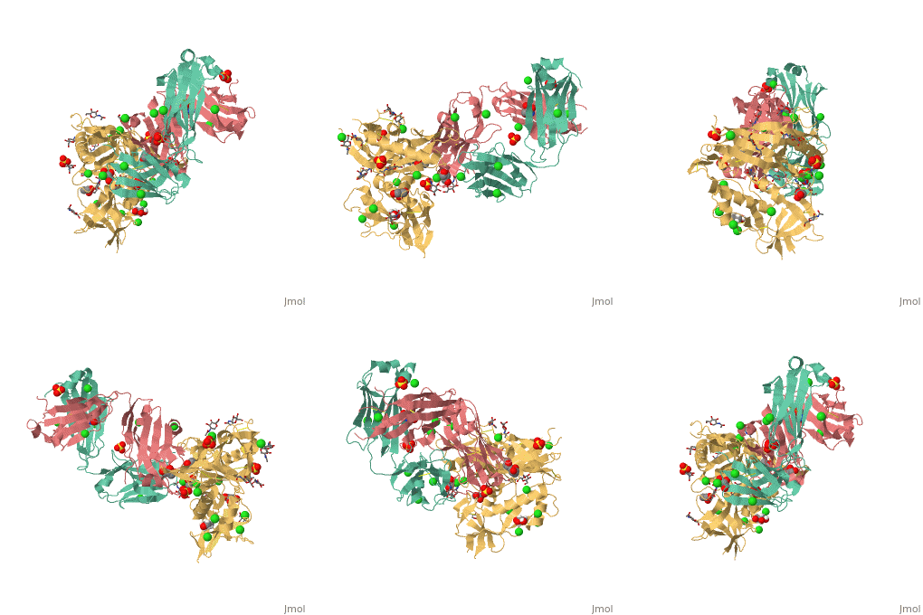
VRC01 gp120 complex
From Proteopedia
| |||||||||||
References
- ↑ 1.0 1.1 1.2 1.3 Wu X, Zhou T, Zhu J, Zhang B, Georgiev I, Wang C, Chen X, Longo NS, Louder M, McKee K, O'Dell S, Perfetto S, Schmidt SD, Shi W, Wu L, Yang Y, Yang ZY, Yang Z, Zhang Z, Bonsignori M, Crump JA, Kapiga SH, Sam NE, Haynes BF, Simek M, Burton DR, Koff WC, Doria-Rose NA, Connors M, Mullikin JC, Nabel GJ, Roederer M, Shapiro L, Kwong PD, Mascola JR. Focused evolution of HIV-1 neutralizing antibodies revealed by structures and deep sequencing. Science. 2011 Sep 16;333(6049):1593-602. Epub 2011 Aug 11. PMID:21835983 doi:10.1126/science.1207532
- ↑ 2.0 2.1 Kwong PD, Mascola JR, Nabel GJ. The changing face of HIV vaccine research. J Int AIDS Soc. 2012 Jul 5;15(2):17407. doi: 10.7448/IAS.15.2.17407. PMID:22789610
- ↑ Li Y, O'Dell S, Walker LM, Wu X, Guenaga J, Feng Y, Schmidt SD, McKee K, Louder MK, Ledgerwood JE, Graham BS, Haynes BF, Burton DR, Wyatt RT, Mascola JR. Mechanism of neutralization by the broadly neutralizing HIV-1 monoclonal antibody VRC01. J Virol. 2011 Sep;85(17):8954-67. Epub 2011 Jun 29. PMID:21715490 doi:10.1128/JVI.00754-11
- ↑ Tran EE, Borgnia MJ, Kuybeda O, Schauder DM, Bartesaghi A, Frank GA, Sapiro G, Milne JL, Subramaniam S. Structural mechanism of trimeric HIV-1 envelope glycoprotein activation. PLoS Pathog. 2012 Jul;8(7):e1002797. Epub 2012 Jul 12. PMID:22807678 doi:10.1371/journal.ppat.1002797
- ↑ 5.0 5.1 5.2 Zhou T, Georgiev I, Wu X, Yang ZY, Dai K, Finzi A, Do Kwon Y, Scheid JF, Shi W, Xu L, Yang Y, Zhu J, Nussenzweig MC, Sodroski J, Shapiro L, Nabel GJ, Mascola JR, Kwong PD. Structural basis for broad and potent neutralization of HIV-1 by antibody VRC01. Science. 2010 Aug 13;329(5993):811-7. Epub 2010 Jul 8. PMID:20616231 doi:10.1126/science.1192819
- ↑ Diskin R, Scheid JF, Marcovecchio PM, West AP Jr, Klein F, Gao H, Gnanapragasam PN, Abadir A, Seaman MS, Nussenzweig MC, Bjorkman PJ. Increasing the Potency and Breadth of an HIV Antibody by Using Structure-Based Rational Design. Science. 2011 Oct 27. PMID:22033520 doi:10.1126/science.1213782
- ↑ 7.0 7.1 Scheid JF, Mouquet H, Ueberheide B, Diskin R, Klein F, Olivera TY, Pietzsch J, Fenyo D, Abadir A, Velinzon K, Hurley A, Myung S, Boulad F, Poignard P, Burton D, Pereyra F, Ho DD, Walker BD, Seaman MS, Bjorkman PJ, Chait BT, Nussenzweig MC. Sequence and Structural Convergence of Broad and Potent HIV Antibodies That Mimic CD4 Binding. Science. 2011 Jul 14. PMID:21764753 doi:10.1126/science.1207227
- ↑ Veazey RS, Shattock RJ, Pope M, Kirijan JC, Jones J, Hu Q, Ketas T, Marx PA, Klasse PJ, Burton DR, Moore JP. Prevention of virus transmission to macaque monkeys by a vaginally applied monoclonal antibody to HIV-1 gp120. Nat Med. 2003 Mar;9(3):343-6. Epub 2003 Feb 10. PMID:12579198 doi:10.1038/nm833
- ↑ Veselinovic M, Neff CP, Mulder LR, Akkina R. Topical gel formulation of broadly neutralizing anti-HIV-1 monoclonal antibody VRC01 confers protection against HIV-1 vaginal challenge in a humanized mouse model. Virology. 2012 Oct 25;432(2):505-10. doi: 10.1016/j.virol.2012.06.025. Epub 2012 , Jul 24. PMID:22832125 doi:10.1016/j.virol.2012.06.025
Proteopedia Page Contributors and Editors (what is this?)
Amanda Valdiosera, Michal Harel, Chris Casey, Alexander Berchansky


