ATPase
From Proteopedia
| Line 3: | Line 3: | ||
[[ATPase]] is an enzyme which catalyzes the breakdown of ATP into ADP and a phosphate ion. This dephosphorylation releases energy which the enzyme uses to drive other reactions. ATPase types include:<br /> | [[ATPase]] is an enzyme which catalyzes the breakdown of ATP into ADP and a phosphate ion. This dephosphorylation releases energy which the enzyme uses to drive other reactions. ATPase types include:<br /> | ||
| - | * '''F-ATPase''' - the prime producers of ATP;<br /> <!--Should there be a F-ATPase page for this like thee is for V-ATPase below? If one is made, I'd like to see links to the animations/movies referenced at https://twitter.com/NathanRoberts17/status/943428752113094656 there.--> | + | * '''F-ATPase''' - the prime producers of ATP. For details see [[Alice Clark/ATPsynthase]];<br /> <!--Should there be a F-ATPase page for this like thee is for V-ATPase below? If one is made, I'd like to see links to the animations/movies referenced at https://twitter.com/NathanRoberts17/status/943428752113094656 there.--> |
* '''V-ATPase''' or Vacuolar-type H+ ATPase couples the energy to proton transport across membranes. For details see [[V-ATPase]];<br /> | * '''V-ATPase''' or Vacuolar-type H+ ATPase couples the energy to proton transport across membranes. For details see [[V-ATPase]];<br /> | ||
* '''A-ATPase''' are found in archaea. For details see [[A-ATP Synthase]];<br /> | * '''A-ATPase''' are found in archaea. For details see [[A-ATP Synthase]];<br /> | ||
Revision as of 08:32, 3 January 2019
| |||||||||||
Contents |
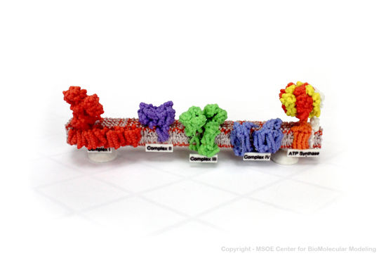
3D Printed Physical Model of ATP Synthase
Shown below is a 3D printed physical model of the Respiration Electron Transport Chain. Complex I is colored red, complex II is purple, complex III is green, complex IV is blue and the atp synthase protein is colored orange, yellow and red.

The MSOE Center for BioMolecular Modeling
The MSOE Center for BioMolecular Modeling uses 3D printing technology to create physical models of protein and molecular structures, making the invisible molecular world more tangible and comprehensible. To view more protein structure models, visit our Model Gallery.
3D Structures of ATPase
Updated on 03-January-2019
V-ATPase
(mutant)
P-ATPase
3ibg - ATPase GET3 – Aspergillus fumigatus
2xit, 2xj4 – CcMipZ – Caulobacter crescentus
2xj9 – CcMipZ (mutant)
References
- ↑ Gorynia S, Bandeiras TM, Pinho FG, McVey CE, Vonrhein C, Round A, Svergun DI, Donner P, Matias PM, Carrondo MA. Structural and functional insights into a dodecameric molecular machine - The RuvBL1/RuvBL2 complex. J Struct Biol. 2011 Sep 10. PMID:21933716 doi:10.1016/j.jsb.2011.09.001
Proteopedia Page Contributors and Editors (what is this?)
Michal Harel, Wayne Decatur, Alexander Berchansky, Mark Hoelzer, Karsten Theis, Jaime Prilusky

