4dpv
From Proteopedia
(Difference between revisions)
(New page: 200px<br /><applet load="4dpv" size="450" color="white" frame="true" align="right" spinBox="true" caption="4dpv, resolution 2.900Å" /> '''PARVOVIRUS/DNA COMP...) |
|||
| (13 intermediate revisions not shown.) | |||
| Line 1: | Line 1: | ||
| - | [[Image:4dpv.gif|left|200px]]<br /><applet load="4dpv" size="450" color="white" frame="true" align="right" spinBox="true" | ||
| - | caption="4dpv, resolution 2.900Å" /> | ||
| - | '''PARVOVIRUS/DNA COMPLEX'''<br /> | ||
| - | == | + | ==PARVOVIRUS/DNA COMPLEX== |
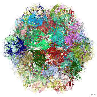
| - | The DNA-containing capsid of canine parvovirus (CPV) is analyzed following | + | <StructureSection load='4dpv' size='340' side='right'caption='[[4dpv]], [[Resolution|resolution]] 2.90Å' scene=''> |
| + | == Structural highlights == | ||
| + | <table><tr><td colspan='2'>[[4dpv]] is a 2 chain structure with sequence from [https://en.wikipedia.org/wiki/Canine_parvovirus Canine parvovirus]. This structure supersedes the now removed PDB entries [http://oca.weizmann.ac.il/oca-bin/send-pdb?obs=1&id=3dpv 3dpv] and [http://oca.weizmann.ac.il/oca-bin/send-pdb?obs=1&id=2dpv 2dpv]. The May 2010 RCSB PDB [https://pdb.rcsb.org/pdb/static.do?p=education_discussion/molecule_of_the_month/index.html Molecule of the Month] feature on ''Parvoviruses'' by David Goodsell is [https://dx.doi.org/10.2210/rcsb_pdb/mom_2010_5 10.2210/rcsb_pdb/mom_2010_5]. Full crystallographic information is available from [http://oca.weizmann.ac.il/oca-bin/ocashort?id=4DPV OCA]. For a <b>guided tour on the structure components</b> use [https://proteopedia.org/fgij/fg.htm?mol=4DPV FirstGlance]. <br> | ||
| + | </td></tr><tr id='ligand'><td class="sblockLbl"><b>[[Ligand|Ligands:]]</b></td><td class="sblockDat" id="ligandDat"><scene name='pdbligand=MG:MAGNESIUM+ION'>MG</scene></td></tr> | ||
| + | <tr id='resources'><td class="sblockLbl"><b>Resources:</b></td><td class="sblockDat"><span class='plainlinks'>[https://proteopedia.org/fgij/fg.htm?mol=4dpv FirstGlance], [http://oca.weizmann.ac.il/oca-bin/ocaids?id=4dpv OCA], [https://pdbe.org/4dpv PDBe], [https://www.rcsb.org/pdb/explore.do?structureId=4dpv RCSB], [https://www.ebi.ac.uk/pdbsum/4dpv PDBsum], [https://prosat.h-its.org/prosat/prosatexe?pdbcode=4dpv ProSAT]</span></td></tr> | ||
| + | </table> | ||
| + | == Function == | ||
| + | [https://www.uniprot.org/uniprot/CAPSD_PAVCD CAPSD_PAVCD] Capsid protein self-assembles to form an icosahedral capsid with a T=1 symmetry, about 22 nm in diameter, and consisting of 60 copies of two size variants of the capsid proteins, VP1 and VP2, which differ by the presence of an N-terminal extension in the minor protein VP1. The capsid encapsulates the genomic ssDNA. Capsid proteins are responsible for the attachment to host cell receptor TFRC. This attachment induces virion internalization predominantly through clathrin-endocytosis. Binding to the host receptors also induces capsid rearrangements leading to surface exposure of VP1 N-terminus, specifically its phospholipase A2-like region and nuclear localization signal(s). VP1 N-terminus might serve as a lipolytic enzyme to breach the endosomal membrane during entry into host cell (By similarity). Intracytoplasmic transport involves microtubules and interaction between capsid proteins and host dynein. Exposure of nuclear localization signal probably allows nuclear import of capsids.<ref>PMID:11799183</ref> <ref>PMID:12970411</ref> <ref>PMID:19656887</ref> | ||
| + | == Evolutionary Conservation == | ||
| + | [[Image:Consurf_key_small.gif|200px|right]] | ||
| + | Check<jmol> | ||
| + | <jmolCheckbox> | ||
| + | <scriptWhenChecked>; select protein; define ~consurf_to_do selected; consurf_initial_scene = true; script "/wiki/ConSurf/dp/4dpv_consurf.spt"</scriptWhenChecked> | ||
| + | <scriptWhenUnchecked>script /wiki/extensions/Proteopedia/spt/initialview01.spt</scriptWhenUnchecked> | ||
| + | <text>to colour the structure by Evolutionary Conservation</text> | ||
| + | </jmolCheckbox> | ||
| + | </jmol>, as determined by [http://consurfdb.tau.ac.il/ ConSurfDB]. You may read the [[Conservation%2C_Evolutionary|explanation]] of the method and the full data available from [http://bental.tau.ac.il/new_ConSurfDB/main_output.php?pdb_ID=4dpv ConSurf]. | ||
| + | <div style="clear:both"></div> | ||
| + | <div style="background-color:#fffaf0;"> | ||
| + | == Publication Abstract from PubMed == | ||
| + | The DNA-containing capsid of canine parvovirus (CPV) is analyzed following atomic refinement at 2.9 A resolution. The capsid contains 60 copies of the capsid protein related by icosahedral symmetry. The atomic model has been extended from the first residue (Gly37) of the unrefined 3.25 A structure towards the N terminus. The electron density shows that approximately 87% of the capsid proteins have N termini on the inside of the capsid, but for approximately 13%, the polypeptide starts on the outside and runs through one of the pores surrounding each 5-fold axis, explaining apparently conflicting antigenic data. Analysis of potential hydrogen bonds reveals approximately 50% more secondary structure than previously apparent. Most of the additional secondary structure are in the 71 and 221 residue-long loop insertions between beta-strands E and F and G and H, forming subunit-bridging sheets that likely add specificity to assembly interactions. Structural analysis of the extensive subunit interactions around the 3-fold axes shows that assembly is a multistep process with loops intertwining following initial contact. Estimated free energies of association suggest that the formation of 3 and 5-fold contacts likely takes precedence over 2-fold interactions. Energies for initial association into trimers or pentamers would be similar, but the intertwining of loops about the 3-fold axis adds an additional large activation barrier to dissociation. Analysis of the surfaces of the assembled capsid shows a surprising lack of basic amino acids that might have been expected to interact with the negatively charged phosphoribose backbone of the DNA. Instead, uncharged polar and van der Waal's interactions predominate in the packaging of single-stranded DNA into the capsid. | ||
| - | + | Canine parvovirus capsid structure, analyzed at 2.9 A resolution.,Xie Q, Chapman MS J Mol Biol. 1996 Dec 6;264(3):497-520. PMID:8969301<ref>PMID:8969301</ref> | |
| - | + | ||
| - | + | From MEDLINE®/PubMed®, a database of the U.S. National Library of Medicine.<br> | |
| - | + | </div> | |
| - | + | <div class="pdbe-citations 4dpv" style="background-color:#fffaf0;"></div> | |
| - | + | ||
| - | + | ||
| - | + | ||
| - | + | ||
| - | + | ||
| - | + | ||
| - | + | ||
| - | + | ||
| - | + | ||
| - | + | ==See Also== | |
| + | *[[Canine parvovirus|Canine parvovirus]] | ||
| + | *[[Virus coat proteins 3D structures|Virus coat proteins 3D structures]] | ||
| + | == References == | ||
| + | <references/> | ||
| + | __TOC__ | ||
| + | </StructureSection> | ||
| + | [[Category: Canine parvovirus]] | ||
| + | [[Category: Large Structures]] | ||
| + | [[Category: Parvoviruses]] | ||
| + | [[Category: RCSB PDB Molecule of the Month]] | ||
| + | [[Category: Chapman MS]] | ||
| + | [[Category: Rossmann MG]] | ||
Current revision
PARVOVIRUS/DNA COMPLEX
| |||||||||||

