Sandbox Home

From Proteopedia

(Difference between revisions)
Jump to: navigation, search
Line 4: Line 4:
<tr>
<tr>
<td colspan="3" style="background:#F5F5FC; border:1px solid #ddd;">
-
<div style="position:relative; top:0.2em; font-size:1.2em; padding:5px 5px 5px 10px; float:right;">
+
<div style="position:relative; top:0.2em; font-size:1.2em; padding:5px 5px 5px 10px; text-align:right; display:block;">
-
<b><i>ISSN 2310-6301</i></b>
+
<b><i>ISSN 2310-6301</i></b>
-
</div>
+
</div>
<span style="display:block; margin:0; padding:0.3em; color:#000; font-style:italic; font-size:1.4em;">

Revision as of 16:13, 30 September 2025

 ISSN 2310-6301
     
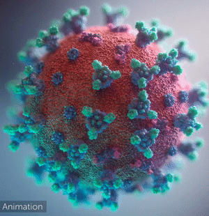
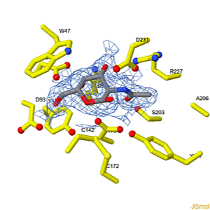
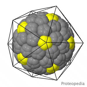
       As life is more than 2D, Proteopedia helps to bridge the gap between 3D structure & function of biomacromolecules
     
     
       Proteopedia presents this information in a user-friendly way as a collaborative & free 3D-encyclopedia of proteins & other biomolecules.
     
Selected Research Pages In Journals Education
About this image
Coronavirus COVID-19

A novel coronavirus was found to be the cause of a respiratory illness first detected in Wuhan, China in 2019. 3D structural studies are aiding scientists to understand how the coronavirus infects humans and helping to find new ways to treat the viral spread (video by Fusion Animation).

>>> Visit this page >>>

About this image
Interconversion of the specificities of human lysosomal enzymes associated with Fabry and Schindler diseases.

IB Tomasic, MC Metcalf, AI Guce, NE Clark, SC Garman. J. Biol. Chem. 2010 doi: 10.1074/jbc.M110.118588
The human lysosomal enzymes α-galactosidase and α-N-acetylgalactosaminidase share 46% amino acid sequence identity and have similar folds. Using a rational protein engineering approach, we interconverted the enzymatic specificity of α-GAL and α-NAGAL. The engineered α-GAL retains the antigenicity but has acquired the enzymatic specificity of α-NAGAL. Conversely, the engineered α-NAGAL retains the antigenicity but has acquired the enzymatic specificity of the α-GAL enzyme. Comparison of the crystal structures of the designed enzyme to the wild-type enzymes shows that active sites superimpose well, indicating success of the rational design. The designed enzymes might be useful as non-immunogenic alternatives in enzyme replacement therapy for treatment of lysosomal storage disorders such as Fabry disease.

>>> Visit this I3DC complement >>>

About this image
Virus Capsid Geometry

The Capsid of a virus is its outer shell or "skin". Viruses have evolved intricate and elegant ways to assemble capsid protein chains into complete, usually spherical capsids, often with icosahedral symmetry. Pictured is an extremely simplified model of a capsid, where a single enlarged atom represents each of the 360 protein chains in the capsid of the Simian Virus 40 (SV40), a member of a group of cancer-causing viruses that has been extensively researched for decades.

>>> See more animations and explanation >>>

About Contact Hot News Table of Contents Structure Index Help
Personal tools