This old version of Proteopedia is provided for student assignments while the new version is undergoing repairs. Content and edits done in this old version of Proteopedia after March 1, 2026 will eventually be lost when it is retired in about June of 2026.
Apply for new accounts at the new Proteopedia. Your logins will work in both the old and new versions.
Cowpea Chlorotic Mottle Virus
From Proteopedia
(Difference between revisions)
| Line 9: | Line 9: | ||
</StructureSection> | </StructureSection> | ||
==3D structures of CPMV== | ==3D structures of CPMV== | ||
| + | Updated on {{REVISIONDAY2}}-{{MONTHNAME|{{REVISIONMONTH}}}}-{{REVISIONYEAR}} | ||
[[1ny7]], [[2bfu]] – CPMV small + large subunits<br /> | [[1ny7]], [[2bfu]] – CPMV small + large subunits<br /> | ||
| - | [[1cwp]] – CPMV coat protein + RNA | + | [[1cwp]] – CPMV coat protein + RNA<br /> |
| + | [[1za7]] – CPMV coat protein (mutant)<br /> | ||
== References == | == References == | ||
<references/> | <references/> | ||
[[Category:Topic Page]] | [[Category:Topic Page]] | ||
Current revision
| |||||||||||
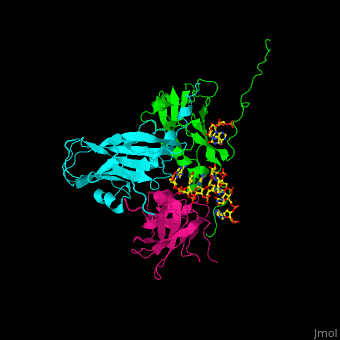
3D structures of CPMV
Updated on 16-May-2019
1ny7, 2bfu – CPMV small + large subunits
1cwp – CPMV coat protein + RNA
1za7 – CPMV coat protein (mutant)
References
- ↑ Speir JA, Munshi S, Wang G, Baker TS, Johnson JE. Structures of the native and swollen forms of cowpea chlorotic mottle virus determined by X-ray crystallography and cryo-electron microscopy. Structure. 1995 Jan 15;3(1):63-78. PMID:7743132
Proteopedia Page Contributors and Editors (what is this?)
Inna Blyakhman, Michal Harel, Alexander Berchansky, Joel L. Sussman
