User:Clayton Moore/Sandbox 1
From Proteopedia
< User:Clayton Moore(Difference between revisions)
| (7 intermediate revisions not shown.) | |||
| Line 2: | Line 2: | ||
== Introduction == | == Introduction == | ||
| - | Hrp1 is a heterogeneous ribonuclear protein of [https://en.wikipedia.org/wiki/Saccharomyces_cerevisiae Saccharomyces cerevisiae], baker’s yeast. Hrp1 is an essential component of 3’ [https://en.wikipedia.org/wiki/Post-transcriptional_modification pre-mRNA processing] and contributes to the preparatory cleavage required for polyadenylation. The gene expressed as Hrp1, HRP1, was first isolated by Henry, et al.<ref> Henry, Michael, et al. “Potential RNA Binding Proteins in Saccharomyces Cerevisiae Identified as Suppressors of Temperature-Sensitive Mutations inNPL3.” Genetics, vol. 142, Jan. 1996, pp. 103–115. </ref> and was later attributed to the Hrp1 protein by Kessler, et al.<ref> Kessler, Marco M, et al. “Purification of the Saccharomyces Cerevisiae Cleavage/Polyadenylation Factor I.” Journal of Biological Chemistry, vol. 271, no. 43, 25 Oct. 1996, pp. 27167–27175. </ref> Hrp1 also participates in the regulation of the 3’ end. | + | __TOC__ |
| - | + | Hrp1 is a heterogeneous ribonuclear protein of [https://en.wikipedia.org/wiki/Saccharomyces_cerevisiae Saccharomyces cerevisiae], baker’s yeast. Hrp1 is an essential component of 3’ [https://en.wikipedia.org/wiki/Post-transcriptional_modification pre-mRNA processing] and contributes to the preparatory cleavage required for polyadenylation. The gene expressed as Hrp1, HRP1, was first isolated by Henry, et al.<ref> Henry, Michael, et al. “Potential RNA Binding Proteins in Saccharomyces Cerevisiae Identified as Suppressors of Temperature-Sensitive Mutations inNPL3.” Genetics, vol. 142, Jan. 1996, pp. 103–115. </ref> and was later attributed to the Hrp1 protein by Kessler, et al.<ref name="Kessler 1996"> Kessler, Marco M, et al. “Purification of the Saccharomyces Cerevisiae Cleavage/Polyadenylation Factor I.” Journal of Biological Chemistry, vol. 271, no. 43, 25 Oct. 1996, pp. 27167–27175. </ref> Hrp1 also participates in the regulation of the 3’ end. The structure was solved via NMR by Pérez-Cañadillas.<ref name="Perez-Canadillas 2006">Pérez-Cañadillas, Jose Manuel. “Grabbing the Message: Structural Basis of MRNA 3â²UTR Recognition by Hrp1.” The EMBO Journal, vol. 25, no. 13, 2006, pp. 3167–3178., doi:10.1038/sj.emboj.7601190. </ref> | |
<StructureSection load='2cjk' size='400' side='right' caption='(PDB entry [[2cjk]])' scene=''> | <StructureSection load='2cjk' size='400' side='right' caption='(PDB entry [[2cjk]])' scene=''> | ||
| Line 10: | Line 10: | ||
=== Polyadenylation Complex === | === Polyadenylation Complex === | ||
| - | Hrp1 is a member of yeast mRNA cleavage factor 1 (CF1), which, along with the cleavage stimulatory factor (CstF), processes the 3’ end, most notably through polyadenylation. Though crucial in eukaryotic pre-mRNA processing, polyadenylation is especially important in yeast, where intron splicing is far less frequent than in higher eukaryotes.<ref> Guisbert, K. Kim. “Functional Specificity of Shuttling HnRNPs Revealed by Genome-Wide Analysis of Their RNA Binding Profiles.” RNA, vol. 11, no. 4, Jan. 2005, pp. 383–393., doi:10.1261/rna.7234205. </ref> CF1 binds mRNA upstream of the cleavage site<ref | + | Hrp1 is a member of yeast mRNA cleavage factor 1 (CF1), which, along with the cleavage stimulatory factor (CstF), processes the 3’ end, most notably through polyadenylation. Though crucial in eukaryotic pre-mRNA processing, polyadenylation is especially important in yeast, where intron splicing is far less frequent than in higher eukaryotes.<ref name="Guisbert 2005"> Guisbert, K. Kim. “Functional Specificity of Shuttling HnRNPs Revealed by Genome-Wide Analysis of Their RNA Binding Profiles.” RNA, vol. 11, no. 4, Jan. 2005, pp. 383–393., doi:10.1261/rna.7234205. </ref> CF1 binds mRNA upstream of the cleavage site<ref name="Kessler 1996" /> and is divided into two components, CF1a and b, the former of which contains Rna14, Rna15, Clp1 and Pfc11.<ref name="Minvielle-Sebastia 1998"> Minvielle-Sebastia, L. “Control of Cleavage Site Selection during MRNA 3 End Formation by a Yeast HnRNP.” The EMBO Journal, vol. 17, no. 24, 1998, pp. 7454–7468., doi:10.1093/emboj/17.24.7454. </ref> Hrp1 is the sole polypeptide of cleavage factor 1b (CF1b).<ref name="Kessler 1997"> Kessler, M. M., et al. “Hrp1, a Sequence-Specific RNA-Binding Protein That Shuttles between the Nucleus and the Cytoplasm, Is Required for MRNA 3-End Formation in Yeast.” Genes & Development, vol. 11, no. 19, Jan. 1997, pp. 2545–2556., doi:10.1101/gad.11.19.2545. </ref> Hrp1 interacts with the CF1a proteins Rna14 and Rna15 in an inverted U-like structure in the presence of mRNA.<ref name="Barnwal 2012"> Barnwal, R. P., et al. “Structural and Biochemical Analysis of the Assembly and Function of the Yeast Pre-MRNA 3 End Processing Complex CF I.” Proceedings of the National Academy of Sciences, vol. 109, no. 52, Oct. 2012, pp. 21342–21347., doi:10.1073/pnas.1214102110.</ref> Rna15 interacts with both RNA recognition motifs (RRMs) of Hrp15<ref name="Barnwal 2012" />, while Rna14 contacts Hrp1 in such a way that maximizes the distance between the negative domains of each protein.<ref> Leeper, Thomas C., et al. “Novel Protein–Protein Contacts Facilitate MRNA 3′-Processing Signal Recognition by Rna15 and Hrp1.” Journal of Molecular Biology, vol. 401, no. 3, 2010, pp. 334–349., doi:10.1016/j.jmb.2010.06.032. </ref> |
=== Specificity and Location=== | === Specificity and Location=== | ||
| - | Yeast hnRNPs, including Hrp1, are specific to certain mRNA strands. Hrp1 is specific to an mRNA efficiency element consisting of alternating UA sequencing<ref | + | Yeast hnRNPs, including Hrp1, are specific to certain mRNA strands. Hrp1 is specific to an mRNA efficiency element consisting of alternating UA sequencing<ref name="Guisbert 2005" /> approximately seven nucleotides in length.<ref> Chen, S. “A Specific RNA-Protein Interaction at Yeast Polyadenylation Efficiency Elements.” Nucleic Acids Research, vol. 26, no. 21, Jan. 1998, pp. 4965–4974., doi:10.1093/nar/26.21.4965. </ref> This sequence specificity, among other observational data, has given credence to the notion, first proposed by Minvielle-Sebastia, et al., that Hrp1 is not totally essential to the 3’ processing of every mRNA.<ref name="Minvielle-Sebastia 1998" /> This notion is further supported by the ability of yeast cells to survive hyperosmotic stress-induced extranuclear export of Hrp1.<ref name="Henry 2003"> Henry, M. F. “The Yeast HnRNP-like Protein Hrp1/Nab4 Accumulates in the Cytoplasm after Hyperosmotic Stress: A Novel Fps1-Dependent Response.” Molecular Biology of the Cell, vol. 14, no. 9, Nov. 2003, pp. 3929–3941., doi:10.1091/mbc.e03-01-0854. </ref> |
| - | Hrp1 participates in mRNA processing within the nucleus, but it may be found endo- or exonuclearly.<ref | + | Hrp1 participates in mRNA processing within the nucleus, but it may be found endo- or exonuclearly.<ref name="Henry 2003" /><ref name="Kessler 1997" /> A nuclear localization signal (NLS) at the C-terminal end of Hrp1 is essential to its recognition by the nuclear transportin receptor Kap104. The receptor and NLS are orthologous to human karyopherin B2 and hnRNP A1.<ref> Lange, Allison, et al. “A PY-NLS Nuclear Targeting Signal Is Required for Nuclear Localization and Function of TheSaccharomyces CerevisiaemRNA-Binding Protein Hrp1.” Journal of Biological Chemistry, vol. 283, no. 19, 2008, pp. 12926–12934., doi:10.1074/jbc.m800898200. </ref> |
=== Regulatory Function === | === Regulatory Function === | ||
| Line 23: | Line 23: | ||
== Structure == | == Structure == | ||
| - | HRP1 is made up of two [https://en.wikipedia.org/wiki/RNA RNA] binding domains (RBDs) that contain residues serving to facilitate RNA recognition. These two domains fold into a βαββαβ [https://en.wikipedia.org/wiki/Protein_secondary_structure secondary structure]<ref>Clery, Antoine, et al. “RNA Recognition Motifs: Boring? Not Quite.” Current Opinion in Structural Biology, Elsevier Current Trends, 30 May 2008, www.sciencedirect.com/science/article/pii/S0959440X08000584.</ref> in an RNA-free environment, allowing Hrp1 to behave rigidly. The <scene name='78/782604/First_rbd/4'>first RBD</scene> includes residues extending from Ser158 to Ala233 and the <scene name='78/782604/Second_rbd/2'>second RBD</scene> extends from Lys244 to Ala318. Both RBDs are composed of a four-stranded [https://en.wikipedia.org/wiki/Beta_sheet beta sheet] with two [https://en.wikipedia.org/wiki/Alpha_helix alpha helices] spanning across one side of the sheet. The linker region is made up of residues spanning from Ile234 to Gly243. When RNA is introduced into the environment, conformational change is demonstrated within the linker region and a <scene name='78/782604/Linker_helix/2'>short two-turn alpha helix</scene> forms from Arg236 to Lys241. The helix that forms is made up of many charged polar residues that [https://en.wikipedia.org/wiki/Salt_bridge_(protein_and_supramolecular) stablilize] themselves through <scene name='78/782604/Salt_bridges/2'>salt bridge interactions</scene> between Arg236-Asp240 and Asp237-Lys241.<ref | + | HRP1 is made up of two [https://en.wikipedia.org/wiki/RNA RNA] binding domains (RBDs) that contain residues serving to facilitate RNA recognition. These two domains fold into a βαββαβ [https://en.wikipedia.org/wiki/Protein_secondary_structure secondary structure]<ref>Clery, Antoine, et al. “RNA Recognition Motifs: Boring? Not Quite.” Current Opinion in Structural Biology, Elsevier Current Trends, 30 May 2008, www.sciencedirect.com/science/article/pii/S0959440X08000584.</ref> in an RNA-free environment, allowing Hrp1 to behave rigidly. The <scene name='78/782604/First_rbd/4'>first RBD</scene> includes residues extending from Ser158 to Ala233 and the <scene name='78/782604/Second_rbd/2'>second RBD</scene> extends from Lys244 to Ala318. Both RBDs are composed of a four-stranded [https://en.wikipedia.org/wiki/Beta_sheet beta sheet] with two [https://en.wikipedia.org/wiki/Alpha_helix alpha helices] spanning across one side of the sheet. The linker region is made up of residues spanning from Ile234 to Gly243. When RNA is introduced into the environment, conformational change is demonstrated within the linker region and a <scene name='78/782604/Linker_helix/2'>short two-turn alpha helix</scene> forms from Arg236 to Lys241. The helix that forms is made up of many charged polar residues that [https://en.wikipedia.org/wiki/Salt_bridge_(protein_and_supramolecular) stablilize] themselves through <scene name='78/782604/Salt_bridges/2'>salt bridge interactions</scene> between Arg236-Asp240 and Asp237-Lys241.<ref name="Perez-Canadillas 2006" /> |
[[Image:Screen Shot 2018-04-02 at 9.57.35 PM.png |150px|left|thumb|'''Figure 1:'''Positively charged cleft within HRP1 in which RNA binds]] | [[Image:Screen Shot 2018-04-02 at 9.57.35 PM.png |150px|left|thumb|'''Figure 1:'''Positively charged cleft within HRP1 in which RNA binds]] | ||
| Line 36: | Line 36: | ||
[[Image:Ade6-Ura7 H-Bonding.png|150px|right|thumb|'''Figure 3:'''Ade6 donating a hydrogen to the 5'O of Ura7.]] | [[Image:Ade6-Ura7 H-Bonding.png|150px|right|thumb|'''Figure 3:'''Ade6 donating a hydrogen to the 5'O of Ura7.]] | ||
=== Adenosine recognition === | === Adenosine recognition === | ||
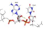
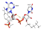
| - | HRP1 specifically binds 3 Adenosine ribonucleotides within the PEE. Adenosine recognition is facilitated through the use of hydrophobic pockets found within HRP1. Ade2 binding is made possible through the interaction of Phe246, which makes up the foundation of the recognition pocket. In addition, the C-terminal of Arg321 interacts with the opposite side of Ade2 through pi-cation interactions. Upon binding, Ade4 is fit inside of a deep hydrophobic pocket made up of Trp168 and Lys226. The stacking of Trp in this interaction is demonstrated as a unique feature of Hrp1; similar Hrp1-like proteins maintain this conserved Trp, but do not demonstrate Trp stacking. The hydrophobic pocket in which Ade6 resides upon binding is made up of Phe162 and Ile234, which sandwich Ade6. In addition, the three Adenosines participating in binding are also recognized by Hydrogen bonds to bases that determine specificity. The three Adenosines recognized display 1, 3, and 2 Hydrogen Bond(s), respectively. The three backbone amides (Glu319 NH, Trp168 NH, Ile234 NH) hydrogen bond with nitrogen atoms of the three adenosine bases (Ade2 N1, Ade4 N7, Ade6 N1). In addition, Ade4 makes base specific contacts with Asn167 and Lys226. Ade4 acts as the donor in it’s interaction with Lys226 and as the acceptor with Asn167. Ade6 interacts with Arg232 in which it acts as the donor. | + | HRP1 specifically binds 3 Adenosine ribonucleotides within the PEE. Adenosine recognition is facilitated through the use of hydrophobic pockets found within HRP1. <scene name='78/782604/Adenosine_2_interactions/1'>Ade2</scene> binding is made possible through the interaction of Phe246, which makes up the foundation of the recognition pocket. In addition, the C-terminal of Arg321 interacts with the opposite side of <scene name='78/782604/Adenosine_2_interactions/1'>Ade2</scene> through pi-cation interactions. Upon binding, <scene name='78/782604/Adenosine_4_interactions/1'>Ade4</scene> is fit inside of a deep hydrophobic pocket made up of Trp168 and Lys226. The stacking of Trp in this interaction is demonstrated as a unique feature of Hrp1; similar Hrp1-like proteins maintain this conserved Trp, but do not demonstrate Trp stacking. The hydrophobic pocket in which <scene name='78/782604/Adenosine_6_interactions/1'>Ade6</scene> resides upon binding is made up of Phe162 and Ile234, which sandwich <scene name='78/782604/Adenosine_6_interactions/1'>Ade6</scene>. In addition, the three Adenosines participating in binding are also recognized by Hydrogen bonds to bases that determine specificity. The three Adenosines recognized display 1, 3, and 2 Hydrogen Bond(s), respectively. The three backbone amides (Glu319 NH, Trp168 NH, Ile234 NH) hydrogen bond with nitrogen atoms of the three adenosine bases (<scene name='78/782604/Adenosine_2_interactions/1'>Ade2</scene> N1, <scene name='78/782604/Adenosine_4_interactions/1'>Ade4</scene> N7, <scene name='78/782604/Adenosine_6_interactions/1'>Ade6</scene> N1). In addition, <scene name='78/782604/Adenosine_4_interactions/1'>Ade4</scene> makes base specific contacts with Asn167 and Lys226. <scene name='78/782604/Adenosine_4_interactions/1'>Ade4</scene> acts as the donor in it’s interaction with Lys226 and as the acceptor with Asn167. <scene name='78/782604/Adenosine_6_interactions/1'>Ade6</scene> interacts with Arg232 in which it acts as the donor. |
| - | <scene name='78/782604/Adenosine_2_interactions/1'> | + | <scene name='78/782604/Adenosine_2_interactions/1'>Ade2</scene> |
| - | <scene name='78/782604/Adenosine_4_interactions/1'> | + | <scene name='78/782604/Adenosine_4_interactions/1'>Ade4</scene> |
| - | <scene name='78/782604/Adenosine_6_interactions/1'> | + | <scene name='78/782604/Adenosine_6_interactions/1'>Ade6</scene> |
=== Uracil recognition === | === Uracil recognition === | ||
| - | Hrp1 interacts with the three uracil bases mainly though Van der Waals contacts. All three uridines interact with aromatic residues of the protein, although Ura3 and Ura5 have low surface accessibility compared to Ura7. Ura3 interacts with Phe288 in a nonplanar position, and Ura5 behaves the same with Phe162. However, Ura7 does form a planar stacking arrangement with the aromatic ring of Phe202. Base discrimination from cytosine relies heavily on the hydrogen bonding between the O4 of the uracil base and the amine group of Lys160. The O2 and O4 of Ura5 also play a role in base discrimination as they form hydrogen bonds with Lys244 and Lys231, respectively. While these two lysines are conserved in most Hrp1-like proteins, there are variations in other organisms that replace Lys244 with an Asn, although the interaction remains conserved. Discrimination at Ura3 is less clear, but it is suggested that it is due to the interaction of its N3 with the backbone phosphate of Ade6. | + | Hrp1 interacts with the three uracil bases mainly though Van der Waals contacts. All three uridines interact with aromatic residues of the protein, although <scene name='78/782604/Uracil_3_interactions/1'>Ura3</scene> and <scene name='78/782604/Uracil_5_interactions/1'>Ura5</scene> have low surface accessibility compared to <scene name='78/782604/Uracil_7_interactions/1'>Ura7</scene>. <scene name='78/782604/Uracil_3_interactions/1'>Ura3</scene> interacts with Phe288 in a nonplanar position, and <scene name='78/782604/Uracil_5_interactions/1'>Ura5</scene> behaves the same with Phe162. However, <scene name='78/782604/Uracil_7_interactions/1'>Ura7</scene> does form a planar stacking arrangement with the aromatic ring of Phe202. Base discrimination from cytosine relies heavily on the hydrogen bonding between the O4 of the uracil base and the amine group of Lys160. The O2 and O4 of <scene name='78/782604/Uracil_5_interactions/1'>Ura5</scene> also play a role in base discrimination as they form hydrogen bonds with Lys244 and Lys231, respectively. While these two lysines are conserved in most Hrp1-like proteins, there are variations in other organisms that replace Lys244 with an Asn, although the interaction remains conserved. Discrimination at <scene name='78/782604/Uracil_3_interactions/1'>Ura3</scene> is less clear, but it is suggested that it is due to the interaction of its N3 with the backbone phosphate of <scene name='78/782604/Adenosine_6_interactions/1'>Ade6</scene>. |
| - | <scene name='78/782604/Uracil_3_interactions/1'> | + | <scene name='78/782604/Uracil_3_interactions/1'>Ura3</scene> |
| - | <scene name='78/782604/Uracil_5_interactions/1'> | + | <scene name='78/782604/Uracil_5_interactions/1'>Ura5</scene> |
| - | <scene name='78/782604/Uracil_7_interactions/1'> | + | <scene name='78/782604/Uracil_7_interactions/1'>Ura7</scene> |
== Novelty == | == Novelty == | ||
| Line 61: | Line 61: | ||
Though Hrp1 is not analogous to any mammalian hnRNP<ref>Gross, S., and C. Moore. “Five Subunits Are Required for Reconstitution of the Cleavage and Polyadenylation Activities of Saccharomyces Cerevisiae Cleavage Factor I.” Proceedings of the National Academy of Sciences, vol. 98, no. 11, Aug. 2001, pp. 6080–6085., doi:10.1073/pnas.101046598.</ref>, the protein and its corresponding gene are occasionally studied as orthologues to human hnRNPs. HNRPDL is one such family of human hnRNPs. Mutations to several members of this class of hnRNPs result in many facets of muscular dystrophy. A study by Vieira, et al. <ref> Vieira, Natássia M., et al. “A Defect in the RNA-Processing Protein HNRPDL Causes Limb-Girdle Muscular Dystrophy 1G (LGMD1G).” Human Molecular Genetics, vol. 23, no. 15, 2014, pp. 4103–4110., doi:10.1093/hmg/ddu127.</ref> found that elimination of Hrp1 had profound effects on protein localization and activation, and these results were used as a model for the genotypic causation of muscular dystrophy. | Though Hrp1 is not analogous to any mammalian hnRNP<ref>Gross, S., and C. Moore. “Five Subunits Are Required for Reconstitution of the Cleavage and Polyadenylation Activities of Saccharomyces Cerevisiae Cleavage Factor I.” Proceedings of the National Academy of Sciences, vol. 98, no. 11, Aug. 2001, pp. 6080–6085., doi:10.1073/pnas.101046598.</ref>, the protein and its corresponding gene are occasionally studied as orthologues to human hnRNPs. HNRPDL is one such family of human hnRNPs. Mutations to several members of this class of hnRNPs result in many facets of muscular dystrophy. A study by Vieira, et al. <ref> Vieira, Natássia M., et al. “A Defect in the RNA-Processing Protein HNRPDL Causes Limb-Girdle Muscular Dystrophy 1G (LGMD1G).” Human Molecular Genetics, vol. 23, no. 15, 2014, pp. 4103–4110., doi:10.1093/hmg/ddu127.</ref> found that elimination of Hrp1 had profound effects on protein localization and activation, and these results were used as a model for the genotypic causation of muscular dystrophy. | ||
| + | |||
| + | Define Hrp's abbreviation better. | ||
| + | Make 2D pictures bigger. | ||
| + | Change Figure 4 colors. | ||
| + | Center RBD 1 image. | ||
| + | Green Link with both RBDs. | ||
| + | Schematic of PolyA Complex | ||
| + | Reorganize Adenosine and Uracil Paragraphs | ||
| + | |||
| + | <scene name='78/782603/Hrp1_and_rna15/1'>Rna 15</scene> | ||
| + | |||
== References == | == References == | ||
<references /> | <references /> | ||
Current revision
HRP1 found in Saccharomyces cerevisiae
Introduction
Contents |
Hrp1 is a heterogeneous ribonuclear protein of Saccharomyces cerevisiae, baker’s yeast. Hrp1 is an essential component of 3’ pre-mRNA processing and contributes to the preparatory cleavage required for polyadenylation. The gene expressed as Hrp1, HRP1, was first isolated by Henry, et al.[1] and was later attributed to the Hrp1 protein by Kessler, et al.[2] Hrp1 also participates in the regulation of the 3’ end. The structure was solved via NMR by Pérez-Cañadillas.[3]
| |||||||||||


