This old version of Proteopedia is provided for student assignments while the new version is undergoing repairs. Content and edits done in this old version of Proteopedia after March 1, 2026 will eventually be lost when it is retired in about June of 2026.
Apply for new accounts at the new Proteopedia. Your logins will work in both the old and new versions.
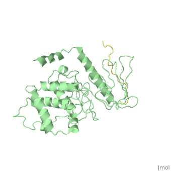
2emt
From Proteopedia
(Difference between revisions)
| Line 3: | Line 3: | ||
<StructureSection load='2emt' size='340' side='right'caption='[[2emt]], [[Resolution|resolution]] 2.80Å' scene=''> | <StructureSection load='2emt' size='340' side='right'caption='[[2emt]], [[Resolution|resolution]] 2.80Å' scene=''> | ||
== Structural highlights == | == Structural highlights == | ||
| - | <table><tr><td colspan='2'>[[2emt]] is a 5 chain structure with sequence from [https://en.wikipedia.org/wiki/ | + | <table><tr><td colspan='2'>[[2emt]] is a 5 chain structure with sequence from [https://en.wikipedia.org/wiki/Mus_musculus Mus musculus]. Full crystallographic information is available from [http://oca.weizmann.ac.il/oca-bin/ocashort?id=2EMT OCA]. For a <b>guided tour on the structure components</b> use [https://proteopedia.org/fgij/fg.htm?mol=2EMT FirstGlance]. <br> |
| - | </td></tr><tr id=' | + | </td></tr><tr id='method'><td class="sblockLbl"><b>[[Empirical_models|Method:]]</b></td><td class="sblockDat" id="methodDat">X-ray diffraction, [[Resolution|Resolution]] 2.8Å</td></tr> |
<tr id='resources'><td class="sblockLbl"><b>Resources:</b></td><td class="sblockDat"><span class='plainlinks'>[https://proteopedia.org/fgij/fg.htm?mol=2emt FirstGlance], [http://oca.weizmann.ac.il/oca-bin/ocaids?id=2emt OCA], [https://pdbe.org/2emt PDBe], [https://www.rcsb.org/pdb/explore.do?structureId=2emt RCSB], [https://www.ebi.ac.uk/pdbsum/2emt PDBsum], [https://prosat.h-its.org/prosat/prosatexe?pdbcode=2emt ProSAT]</span></td></tr> | <tr id='resources'><td class="sblockLbl"><b>Resources:</b></td><td class="sblockDat"><span class='plainlinks'>[https://proteopedia.org/fgij/fg.htm?mol=2emt FirstGlance], [http://oca.weizmann.ac.il/oca-bin/ocaids?id=2emt OCA], [https://pdbe.org/2emt PDBe], [https://www.rcsb.org/pdb/explore.do?structureId=2emt RCSB], [https://www.ebi.ac.uk/pdbsum/2emt PDBsum], [https://prosat.h-its.org/prosat/prosatexe?pdbcode=2emt ProSAT]</span></td></tr> | ||
</table> | </table> | ||
== Function == | == Function == | ||
| - | + | [https://www.uniprot.org/uniprot/RADI_MOUSE RADI_MOUSE] Probably plays a crucial role in the binding of the barbed end of actin filaments to the plasma membrane. | |
== Evolutionary Conservation == | == Evolutionary Conservation == | ||
[[Image:Consurf_key_small.gif|200px|right]] | [[Image:Consurf_key_small.gif|200px|right]] | ||
| Line 36: | Line 36: | ||
</StructureSection> | </StructureSection> | ||
[[Category: Large Structures]] | [[Category: Large Structures]] | ||
| - | [[Category: | + | [[Category: Mus musculus]] |
| - | [[Category: Hakoshima | + | [[Category: Hakoshima T]] |
| - | [[Category: Kitano | + | [[Category: Kitano K]] |
| - | [[Category: Maesaki | + | [[Category: Maesaki R]] |
| - | [[Category: Takai | + | [[Category: Takai Y]] |
| - | [[Category: Terawaki | + | [[Category: Terawaki S]] |
| - | + | ||
| - | + | ||
Current revision
Crystal Structure Analysis of the radixin FERM domain complexed with adhesion molecule PSGL-1
| |||||||||||