This old version of Proteopedia is provided for student assignments while the new version is undergoing repairs. Content and edits done in this old version of Proteopedia after March 1, 2026 will eventually be lost when it is retired in about June of 2026.
Apply for new accounts at the new Proteopedia. Your logins will work in both the old and new versions.
3atg
From Proteopedia
(Difference between revisions)
| (One intermediate revision not shown.) | |||
| Line 4: | Line 4: | ||
== Structural highlights == | == Structural highlights == | ||
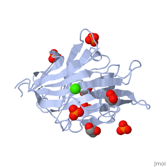
<table><tr><td colspan='2'>[[3atg]] is a 1 chain structure with sequence from [https://en.wikipedia.org/wiki/Cellulosimicrobium_cellulans Cellulosimicrobium cellulans]. Full crystallographic information is available from [http://oca.weizmann.ac.il/oca-bin/ocashort?id=3ATG OCA]. For a <b>guided tour on the structure components</b> use [https://proteopedia.org/fgij/fg.htm?mol=3ATG FirstGlance]. <br> | <table><tr><td colspan='2'>[[3atg]] is a 1 chain structure with sequence from [https://en.wikipedia.org/wiki/Cellulosimicrobium_cellulans Cellulosimicrobium cellulans]. Full crystallographic information is available from [http://oca.weizmann.ac.il/oca-bin/ocashort?id=3ATG OCA]. For a <b>guided tour on the structure components</b> use [https://proteopedia.org/fgij/fg.htm?mol=3ATG FirstGlance]. <br> | ||
| - | </td></tr><tr id='ligand'><td class="sblockLbl"><b>[[Ligand|Ligands:]]</b></td><td class="sblockDat" id="ligandDat"><scene name='pdbligand=CA:CALCIUM+ION'>CA</scene>, <scene name='pdbligand=GOL:GLYCEROL'>GOL</scene>, <scene name='pdbligand=PO4:PHOSPHATE+ION'>PO4</scene | + | </td></tr><tr id='method'><td class="sblockLbl"><b>[[Empirical_models|Method:]]</b></td><td class="sblockDat" id="methodDat">X-ray diffraction, [[Resolution|Resolution]] 1.66Å</td></tr> |
| - | + | <tr id='ligand'><td class="sblockLbl"><b>[[Ligand|Ligands:]]</b></td><td class="sblockDat" id="ligandDat"><scene name='pdbligand=CA:CALCIUM+ION'>CA</scene>, <scene name='pdbligand=GOL:GLYCEROL'>GOL</scene>, <scene name='pdbligand=PO4:PHOSPHATE+ION'>PO4</scene></td></tr> | |
<tr id='resources'><td class="sblockLbl"><b>Resources:</b></td><td class="sblockDat"><span class='plainlinks'>[https://proteopedia.org/fgij/fg.htm?mol=3atg FirstGlance], [http://oca.weizmann.ac.il/oca-bin/ocaids?id=3atg OCA], [https://pdbe.org/3atg PDBe], [https://www.rcsb.org/pdb/explore.do?structureId=3atg RCSB], [https://www.ebi.ac.uk/pdbsum/3atg PDBsum], [https://prosat.h-its.org/prosat/prosatexe?pdbcode=3atg ProSAT]</span></td></tr> | <tr id='resources'><td class="sblockLbl"><b>Resources:</b></td><td class="sblockDat"><span class='plainlinks'>[https://proteopedia.org/fgij/fg.htm?mol=3atg FirstGlance], [http://oca.weizmann.ac.il/oca-bin/ocaids?id=3atg OCA], [https://pdbe.org/3atg PDBe], [https://www.rcsb.org/pdb/explore.do?structureId=3atg RCSB], [https://www.ebi.ac.uk/pdbsum/3atg PDBsum], [https://prosat.h-its.org/prosat/prosatexe?pdbcode=3atg ProSAT]</span></td></tr> | ||
</table> | </table> | ||
| - | + | == Function == | |
| - | = | + | [https://www.uniprot.org/uniprot/G1ED17_CELCE G1ED17_CELCE] |
| - | + | ||
| - | + | ||
| - | + | ||
| - | + | ||
| - | + | ||
| - | + | ||
| - | + | ||
==See Also== | ==See Also== | ||
*[[Glucanase|Glucanase]] | *[[Glucanase|Glucanase]] | ||
*[[Glucanase 3D structures|Glucanase 3D structures]] | *[[Glucanase 3D structures|Glucanase 3D structures]] | ||
| - | == References == | ||
| - | <references/> | ||
__TOC__ | __TOC__ | ||
</StructureSection> | </StructureSection> | ||
[[Category: Cellulosimicrobium cellulans]] | [[Category: Cellulosimicrobium cellulans]] | ||
| - | [[Category: Glucan endo-1,3-beta-D-glucosidase]] | ||
[[Category: Large Structures]] | [[Category: Large Structures]] | ||
| - | [[Category: Mikami | + | [[Category: Mikami B]] |
| - | [[Category: Oda | + | [[Category: Oda M]] |
| - | [[Category: Pang | + | [[Category: Pang Z]] |
| - | [[Category: Tanabe | + | [[Category: Tanabe Y]] |
| - | + | ||
| - | + | ||
| - | + | ||
Current revision
endo-1,3-beta-glucanase from Cellulosimicrobium cellulans
| |||||||||||