We apologize for Proteopedia being slow to respond. For the past two years, a new implementation of Proteopedia has been being built. Soon, it will replace this 18-year old system. All existing content will be moved to the new system at a date that will be announced here.
Lac repressor
From Proteopedia
(Difference between revisions)
| (3 intermediate revisions not shown.) | |||
| Line 1: | Line 1: | ||
__NOTOC__ | __NOTOC__ | ||
| - | <StructureSection load=' | + | <StructureSection load='' size='375' side='right' scene='Morphs/1osl_19_1l1m_9_morph/2' caption=''> |
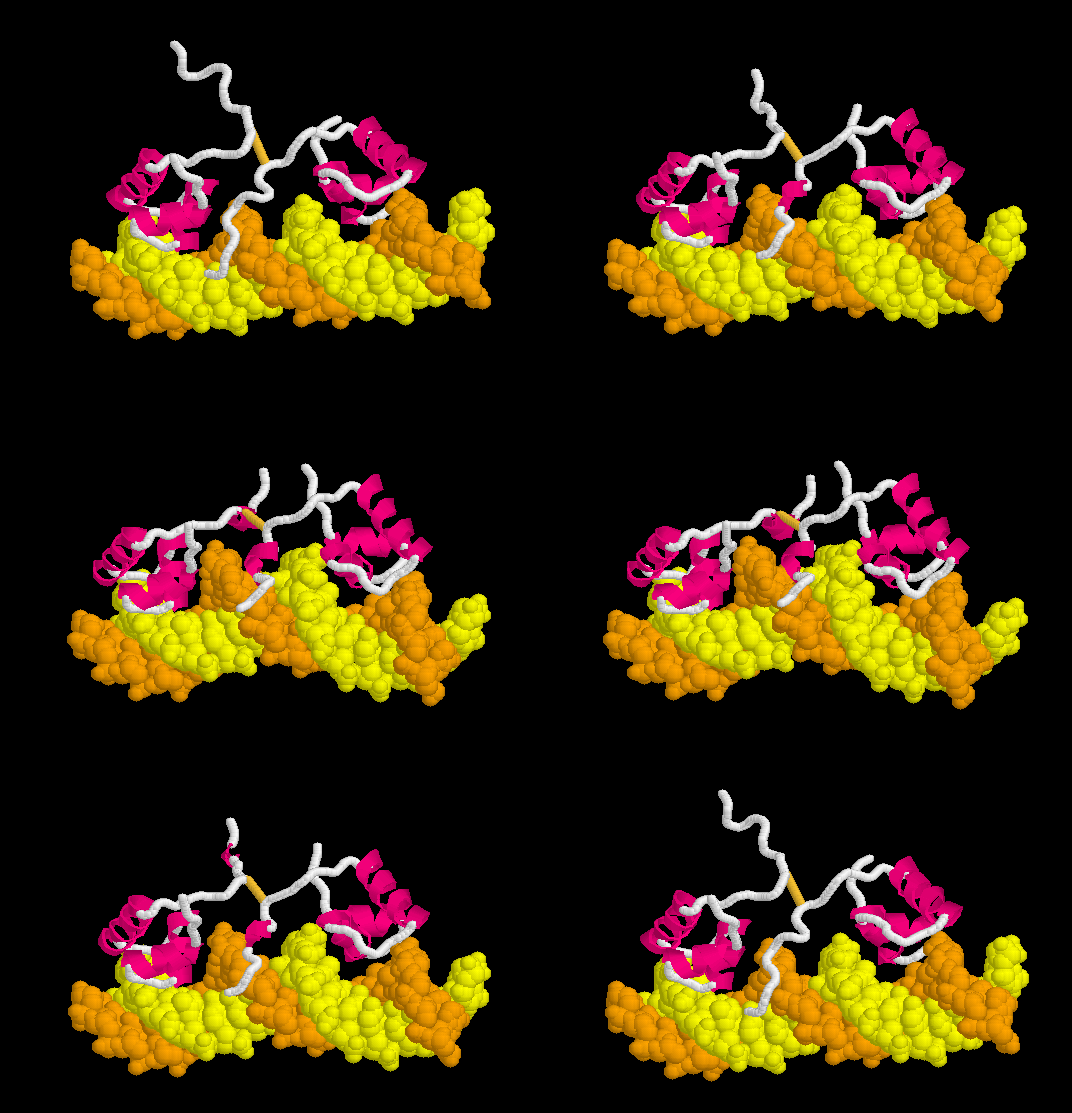
[[Morphs|Morph]] of the '''lac repressor''' complexed with DNA | [[Morphs|Morph]] of the '''lac repressor''' complexed with DNA | ||
| - | <scene name="Morphs/1osl_19_1l1m_9_morph/2">restore initial scene</scene> {{Template:Button Toggle Animation2}} | + | (<scene name="Morphs/1osl_19_1l1m_9_morph/2">restore initial scene</scene>) After displaying interactive model: {{Template:Button Toggle Animation2}} |
showing the differences between non-specific binding (straight DNA) vs. specific recognition of the operator sequence (kinked DNA). Whether the binding kinks the DNA, or simply stabilizes a pre-existing kink, is unknown. [[#Specific Binding| Details Below]]. | showing the differences between non-specific binding (straight DNA) vs. specific recognition of the operator sequence (kinked DNA). Whether the binding kinks the DNA, or simply stabilizes a pre-existing kink, is unknown. [[#Specific Binding| Details Below]]. | ||
__TOC__ | __TOC__ | ||
| Line 50: | Line 50: | ||
====DNA Kinks==== | ====DNA Kinks==== | ||
| - | Strictly speaking, ''bends'' in DNA are distinguished from ''kinks''. DNA is said to be '''kinked''' when the stacking contact between two adjacent base pairs is disrupted<ref name="rohsrev2010" />. The DNA on either side of a kink may be straight or bent. A <scene name='Lac_repressor/Kink/2'>kink occurs in the complex between the lac repressor and specific DNA</scene>: a single CpG base pair is partially separated from the adjacent CpG base pair. <scene name='Lac_repressor/Kink/3'>Zoom in</scene>. Pyrimidine-purine base pairs have the weakest stacking interactions, and are most susceptible to kinking<ref name="rohsrev2010" />. In the complex of lac repressor with specific DNA, <scene name='Lac_repressor/Kink_leu56/1'>two leucines (Leu56)</scene> (<jmol> | + | Strictly speaking, ''bends'' in DNA are distinguished from ''kinks''. DNA is said to be '''kinked''' when the stacking contact between two adjacent base pairs is disrupted<ref name="rohsrev2010" />. The DNA on either side of a kink may be straight or bent. A <scene name='Lac_repressor/Kink/2'>kink occurs in the complex between the lac repressor and specific DNA</scene>: a single CpG base pair is partially separated from the adjacent CpG base pair. <scene name='Lac_repressor/Kink/3'>Zoom in</scene>. Pyrimidine-purine base pairs have the weakest stacking interactions, and are most susceptible to kinking<ref name="rohsrev2010" />. In the complex of lac repressor with specific DNA, <scene name='Lac_repressor/Kink_leu56/1'>two leucines (Leu56)</scene> (if scene is blank,<jmol> |
<jmolLink> | <jmolLink> | ||
<script> model 0;</script> | <script> model 0;</script> | ||
| - | <text>click | + | <text>please click</text> |
</jmolLink> | </jmolLink> | ||
</jmol>)<!--(<font color="red">Sorry, this scene is temporarily broken.</font>)--> are partially intercalated between the separated CpG base pairs, which helps to stabilize the kink. It may often be the case that sequence-dependent kinks and bends are present in DNA prior to the binding of protein<ref name="rohsrev2010" />. DNA structure is dynamic. For example, recently Hoogsteen base pairing was observed to occur transiently in equilibrium with Watson-Crick base pairing<ref>PMID: 21270796</ref> (See ''News & Views''<ref>PMID: 21350476</ref>). Also, the binding of p53 to some but not all DNA sequences stabilizes Hoogsteen (rather than Watson-Crick) base pairing<ref>PMID: 20364130</ref>. Thus, the "bending" (actually kinking) depicted in '''the morph on this page may give the wrong impression''': lac repressor binding may simply stabilize a kink (or transient kink) that pre-existed in the cognate DNA sequence. | </jmol>)<!--(<font color="red">Sorry, this scene is temporarily broken.</font>)--> are partially intercalated between the separated CpG base pairs, which helps to stabilize the kink. It may often be the case that sequence-dependent kinks and bends are present in DNA prior to the binding of protein<ref name="rohsrev2010" />. DNA structure is dynamic. For example, recently Hoogsteen base pairing was observed to occur transiently in equilibrium with Watson-Crick base pairing<ref>PMID: 21270796</ref> (See ''News & Views''<ref>PMID: 21350476</ref>). Also, the binding of p53 to some but not all DNA sequences stabilizes Hoogsteen (rather than Watson-Crick) base pairing<ref>PMID: 20364130</ref>. Thus, the "bending" (actually kinking) depicted in '''the morph on this page may give the wrong impression''': lac repressor binding may simply stabilize a kink (or transient kink) that pre-existed in the cognate DNA sequence. | ||
Current revision
| |||||||||||
3D structures of Lac repressor
Updated on 06-February-2025
See Also
- DNA-protein interactions, an overview introducing helix-turn-helix, leucine zipper, and zinc finger proteins.
- Category: Lac repressor and Category: Lac Repressor, automatically-generated pages that list PDB codes for lac repressor models.
- Morphs where the morph of the lac repressor is used as an example.
- Lac repressor morph methods
- See: Regulation of Gene Expression for additional mechanisms of Gene Regulation
- For additional information, see: Transcription and RNA Processing
References & Notes
- ↑ L'opéron: groupe de gènes à expression coordonée par un opérateur. [Operon: a group of genes with the expression coordinated by an operator.] C R Hebd Seances Acad Sci., 250:1727-9, 1960. PubMed 14406329
- ↑ The lac repressor. Lewis, M. C R Biol. 328:521-48, 2005. PubMed 15950160
- ↑ This domain coloring scheme is adapted from Fig. 6 in the review by Lewis (C. R. Biol. 328:521, 2005). Domains are 1-45, 46-62, (63-162,291-320), (163-290,321-332), 330-339, and 340-357.
- ↑ Conservation results for 1lbg are from the precalculated ConSurf Database, using 103 sequences from Swiss-Prot with an average pairwise distance of 2.4.
- ↑ Conservation results for 1lbi are from the ConSurf Server, using 100 sequences from Uniprot with an average pairwise distance of 1.3.
- ↑ 6.0 6.1 For these scenes, the 20-model PDB files for 1osl and 1l1m were reduced in size, to avoid exceeding the java memory available to the Jmol applet. All atoms except amino acid alpha carbons and DNA phosphorus atoms were removed using the free program alphac.exe from PDBTools. Secondary structure HELIX records from the original PDB file header were retained. The results are Image:1osl ca.pdb and Image:1l1m ca.pdb.
- ↑ Hammar P, Leroy P, Mahmutovic A, Marklund EG, Berg OG, Elf J. The lac repressor displays facilitated diffusion in living cells. Science. 2012 Jun 22;336(6088):1595-8. PMID:22723426 doi:10.1126/science.1221648
- ↑ 8.0 8.1 8.2 8.3 8.4 8.5 8.6 Rohs R, Jin X, West SM, Joshi R, Honig B, Mann RS. Origins of specificity in protein-DNA recognition. Annu Rev Biochem. 2010;79:233-69. PMID:20334529 doi:10.1146/annurev-biochem-060408-091030
- ↑ Joshi R, Passner JM, Rohs R, Jain R, Sosinsky A, Crickmore MA, Jacob V, Aggarwal AK, Honig B, Mann RS. Functional specificity of a Hox protein mediated by the recognition of minor groove structure. Cell. 2007 Nov 2;131(3):530-43. PMID:17981120 doi:10.1016/j.cell.2007.09.024
- ↑ 10.0 10.1 10.2 Rohs R, West SM, Sosinsky A, Liu P, Mann RS, Honig B. The role of DNA shape in protein-DNA recognition. Nature. 2009 Oct 29;461(7268):1248-53. PMID:19865164 doi:10.1038/nature08473
- ↑ Nikolova EN, Kim E, Wise AA, O'Brien PJ, Andricioaei I, Al-Hashimi HM. Transient Hoogsteen base pairs in canonical duplex DNA. Nature. 2011 Feb 24;470(7335):498-502. Epub 2011 Jan 26. PMID:21270796 doi:10.1038/nature09775
- ↑ Honig B, Rohs R. Biophysics: Flipping Watson and Crick. Nature. 2011 Feb 24;470(7335):472-3. PMID:21350476 doi:10.1038/470472a
- ↑ Kitayner M, Rozenberg H, Rohs R, Suad O, Rabinovich D, Honig B, Shakked Z. Diversity in DNA recognition by p53 revealed by crystal structures with Hoogsteen base pairs. Nat Struct Mol Biol. 2010 Apr;17(4):423-9. Epub 2010 Apr 4. PMID:20364130 doi:10.1038/nsmb.1800
- ↑ Powerpoint is a registered trademark for a software package licensed by Microsoft Corp..
Proteopedia Page Contributors and Editors (what is this?)
Eric Martz, Michal Harel, Alexander Berchansky, Joel L. Sussman, Karsten Theis, Henry Jakubowski, David Canner, Eran Hodis, Jaime Prilusky


