Journal:Acta Cryst D:S2059798322009755
From Proteopedia
(Difference between revisions)

| (4 intermediate revisions not shown.) | |||
| Line 12: | Line 12: | ||
<scene name='93/931041/Cv1/6'>Comparison of the human AAR2Δloop-PRPF8RH complex and the yeast Aar2pΔloop-Prp8pRH-Prp8pJM complex</scene> (PDB ID [[4ilg]])<ref name='Weber'>PMID: 23442228</ref>. | <scene name='93/931041/Cv1/6'>Comparison of the human AAR2Δloop-PRPF8RH complex and the yeast Aar2pΔloop-Prp8pRH-Prp8pJM complex</scene> (PDB ID [[4ilg]])<ref name='Weber'>PMID: 23442228</ref>. | ||
| - | <scene name='93/931041/Cv1/7'>Close-up view of interface region I of the yeast Aar2pΔloop-Prp8pRH-Prp8pJM complex</scene>. Water molecules are shown as red spheres. | + | <scene name='93/931041/Cv1/7'>Close-up view of interface region I of the yeast Aar2pΔloop-Prp8pRH-Prp8pJM complex</scene>. <b><span class="text-red">Water molecules are shown as red spheres</span></b>. Interacting residues are shown as ball-and-sticks colored by atom type; carbon, as the respective protein; <b><span class="text-blue">nitrogen, blue</span></b>; <b><span class="text-red">oxygen, red</span></b>; <span class="bg-yellow">sulfur, yellow</span>; dashed black lines, hydrogen bonds or salt bridges. |
<scene name='93/931041/Cv1/8'>Close-up view of interface region I of the human AAR2Δloop-PRPF8RH complex</scene>. | <scene name='93/931041/Cv1/8'>Close-up view of interface region I of the human AAR2Δloop-PRPF8RH complex</scene>. | ||
| Line 20: | Line 20: | ||
<scene name='93/931041/Cv1/11'>Close-up view of interface region II of the human AAR2Δloop-PRPF8RH complex</scene>. | <scene name='93/931041/Cv1/11'>Close-up view of interface region II of the human AAR2Δloop-PRPF8RH complex</scene>. | ||
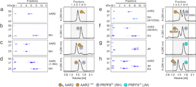
| - | [[Image:Fig2AAR2.png|left|400px|thumb|Figure 2. Probing | + | [[Image:Fig2AAR2.png|left|400px|thumb|Figure 2. Probing AAR2<sup>Δloop</sup>-PRPF8<sup>RH</sup> interacting regions and residues. (a-h) SDS-PAGE analyses (left) and UV elution profiles (right) of analytical size exclusion chromatography runs monitoring the interactions among AAR2 variants, PRPF8<sup>RH</sup> variants and PRPF8<sup>JM</sup>. Figures '''a-c''' were adapted from (Santos et al., 2015)<ref name='Santos'>PMID: 26527271</ref> and are shown for comparison. M, molecular mass standard (kDa); I, input samples. Protein bands are identified on the right. Elution fractions are indicated at the top of the gels and profiles, elution volumes are indicated at the bottom of the profiles. Icons are explained at the bottom. Variants are indicated at the respective icons. Peaks labeled by transparent icons represent an excess of the respective protein.]] |
{{Clear}} | {{Clear}} | ||
| Line 32: | Line 32: | ||
*<scene name='93/931041/Cv/20'>observed in the AAR2Δloop-PRPF8RH complex</scene>; | *<scene name='93/931041/Cv/20'>observed in the AAR2Δloop-PRPF8RH complex</scene>; | ||
*<scene name='93/931041/Cv/21'>modeled onto the PRPF8RH domain in step 1 conformation</scene> (PDB ID [[4jk7]]<ref name='Schellenberg'>PMID: 23686287</ref>); | *<scene name='93/931041/Cv/21'>modeled onto the PRPF8RH domain in step 1 conformation</scene> (PDB ID [[4jk7]]<ref name='Schellenberg'>PMID: 23686287</ref>); | ||
| - | *<scene name='93/931041/Cv/23'>modeled onto the PRPF8RH domain in step 2 conformation</scene> (PDB ID [[4jk7]]). Yellow sphere, coordinated Mg2+ ion. The AAR2 C-terminus clashes with the | + | *<scene name='93/931041/Cv/23'>modeled onto the PRPF8RH domain in step 2 conformation</scene> (PDB ID [[4jk7]]). <span class="bg-yellow">Yellow sphere, coordinated Mg2+ ion</span>. The AAR2 C-terminus clashes with the PRPF8<sup>RH</sup> domain the step 2 conformation. |
The structure and functional data of the human spliceosomal assembly factor Aar2 in complex with a core spliceosomal domain of the PRPF8 protein indicates a different function of human Aar2 in contrast to the yeast protein. | The structure and functional data of the human spliceosomal assembly factor Aar2 in complex with a core spliceosomal domain of the PRPF8 protein indicates a different function of human Aar2 in contrast to the yeast protein. | ||
Current revision
| |||||||||||
This page complements a publication in scientific journals and is one of the Proteopedia's Interactive 3D Complement pages. For aditional details please see I3DC.


