Dihydrofolate reductase
From Proteopedia
| Line 1: | Line 1: | ||
[[Image:1rx2.jpg|left|150px]]<br /> | [[Image:1rx2.jpg|left|150px]]<br /> | ||
| - | <applet load='1rx2' size=' | + | <applet load='1rx2' size='450' color='white' frame='true' spin='on' caption='DHFR' align='right' /> |
'''3D structure of dihydrofolate reductase'''<br /> | '''3D structure of dihydrofolate reductase'''<br /> | ||
== The sole source of tetrahydrofolate== | == The sole source of tetrahydrofolate== | ||
Revision as of 07:49, 10 May 2008
|
3D structure of dihydrofolate reductase
The sole source of tetrahydrofolate
DHFR is a ubiquitous enzyme found in all organisms. The primary physiological role of DHFR is maintenance of the intracellular levels of tetrahydrofolate, a precursor of cofactors required for the biosynthesis of purines, pyrimidines, and several amino acids.
The enzyme, which is the sole source of tetrahydrofolate catalyzes the reduction of (DHF) to 5,6,7,8-tetrahydrofolate (THF) by stereospecific hydride transfer from the cofactor to the C6 atom of the pterin ring with concomitant protonation at N5. Being the sole source of THF it is an Achilles’ heel of rapidly proliferating cells, making it an attractive target of several important anticancer and antimicrobial drugs such as methotrexate, trimethoprim, and pyrimethamine.
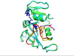
E. coli DHFR is a small (18 kD) protein with an α/β structure consisting of a central eight-stranded β-sheet and four flanking α-helices. The active site cleft divides the protein into two structural subdomains: the (red) and the (green). The adenosine binding subdomain is the smaller of the two subdomains and provides the binding site for the adenosine moiety of the cofactor. The major subdomain consists of ~100 residues from the N and C termini and is dominated by a set of three loops on the ligand binding face that surround the active site. In terms of sequence length, these loops make up approximately 40%–50% of the major subdomain; hence it is sometimes called the “loop” subdomain. The loops are termed Met20, F-G, and G-H loops. Hinge bending motions about Lys38 and Val88 allow the adenosine binding domain to move relative to the major domain upon binding of various ligands, resulting in closure of the active site cleft.
A movie depicting the conformational changes of DHFR during one turnover of substrate has been constructed by M. R. Sawaya and J. Kraut using six isomorphous crystallographic structures. http://chem-faculty.ucsd.edu/kraut/dhfr.html
Proteopedia Page Contributors and Editors (what is this?)
Michal Harel, Karsten Theis, Alexander Berchansky, Joel L. Sussman, Tzvia Selzer, Jaime Prilusky, Eric Martz, Eran Hodis, David Canner

