Sandbox Reserved 641
From Proteopedia
(→'''Structure''') |
(→'''Structure''') |
||
| Line 10: | Line 10: | ||
== '''Structure''' == | == '''Structure''' == | ||
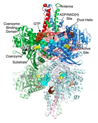
| - | Glutamate Dehydrogenase is a hexamer that is comprised of two trimer subunits. These two subunits are stacked on top of each other and composed of three domains. The top of each domain contains a "NAD-binding domain" that has the conserved nucleotide-binding motif. A larger helix-loop-helix structure rises above this and is referred to as an "antenna." This antenna contains approximately 50 amino acids and is thought to play a major role in regulation of the enzyme. This antennae structure is only found in animals. The bottom domain contacts a domain in the other trimer, | + | Glutamate Dehydrogenase is a hexamer that is comprised of two trimer subunits. These two subunits are stacked on top of each other and composed of three domains. The top of each domain contains a "NAD-binding domain" that has the conserved nucleotide-binding motif. A larger helix-loop-helix structure rises above this and is referred to as an "antenna." This antenna contains approximately 50 amino acids and is thought to play a major role in regulation of the enzyme. This antennae structure is only found in animals. The bottom domain contacts a domain in the other trimer, holding the two trimers together. |
| + | When a substrate binds to the enzyme it binds to the deep recess of the cleft between the NAD binding domain and the lower domain. Along the outside surface of the cleft a coenzyme binds causing the binding domain to rotate by about 18 degrees and close down on the substrate and coenzyme. | ||
| + | |||
| + | Substrate binds to the deep recesses of the cleft between the NAD binding domain and the lower domain. | ||
| + | Coenzyme binds along the NAD binding domain surface of the | ||
| + | cleft. Upon binding, the NAD binding domain rotates by �188 to | ||
| + | firmly close down upon the substrate and coenzyme. As the | ||
| + | catalytic cleft closes, the base of each of the long ascending helices | ||
| + | in the antenna appears to rotate out in a counter-clockwisemanner | ||
| + | to push against the ‘pivot’ helix of the adjacent subunit. There is a | ||
| + | short helix in the descending loop of the antenna that becomes | ||
| + | distended as the mouth closes in a manner akin to an extending | ||
| + | spring. The ‘pivot helix’ rotates in a counter clockwise manner | ||
| + | along the helical axes as well as rotating counter clockwise around | ||
| + | the trimer 3-fold axis. Finally, the entire hexamer seems to ‘exhale’, | ||
| + | or compress, as the mouth closes. This compression is where the | ||
| + | three stacked dimers draw closer to each other, drawing the 2-fold | ||
| + | related subunits closer and compressing the inner core. Therefore, | ||
| + | it is clear that the conformational changes associated with, and | ||
| + | necessary for, catalysis involve the entire hexamer. This not only | ||
| + | might explain the complex kinetic behavior such as negative | ||
| + | cooperativity, but also creates a number of potential binding sites | ||
| + | for allosteric regulators. | ||
[[Image:structure.jpeg]] | [[Image:structure.jpeg]] | ||
Revision as of 01:12, 8 November 2012
| This Sandbox is Reserved from 30/08/2012, through 01/02/2013 for use in the course "Proteins and Molecular Mechanisms" taught by Robert B. Rose at the North Carolina State University, Raleigh, NC USA. This reservation includes Sandbox Reserved 636 through Sandbox Reserved 685. | |||||||
To get started:
More help: Help:Editing For more help, look at this link: http://proteopedia.org/w/Help:Getting_Started_in_Proteopedia
Glutamate Dehydrogenase
IntroductionGlutamate Dehydrogenase (GDH)is used to remove the ketone group and replace it with an α-amine group on the α-carbon, which forms glutaamte. Glutamate is one of the 20 essential amino acids. This is done in reverse to supply α-ketoglutarate to the tricarboxylic acid (TCA) cycle. GDH is an oxidoreductase, which is an enzyme that transfers electrons from one molecule (reductant/electron donor) to another molecule (oxidant/electron acceptor). StructureGlutamate Dehydrogenase is a hexamer that is comprised of two trimer subunits. These two subunits are stacked on top of each other and composed of three domains. The top of each domain contains a "NAD-binding domain" that has the conserved nucleotide-binding motif. A larger helix-loop-helix structure rises above this and is referred to as an "antenna." This antenna contains approximately 50 amino acids and is thought to play a major role in regulation of the enzyme. This antennae structure is only found in animals. The bottom domain contacts a domain in the other trimer, holding the two trimers together. When a substrate binds to the enzyme it binds to the deep recess of the cleft between the NAD binding domain and the lower domain. Along the outside surface of the cleft a coenzyme binds causing the binding domain to rotate by about 18 degrees and close down on the substrate and coenzyme. Substrate binds to the deep recesses of the cleft between the NAD binding domain and the lower domain. Coenzyme binds along the NAD binding domain surface of the cleft. Upon binding, the NAD binding domain rotates by �188 to firmly close down upon the substrate and coenzyme. As the catalytic cleft closes, the base of each of the long ascending helices in the antenna appears to rotate out in a counter-clockwisemanner to push against the ‘pivot’ helix of the adjacent subunit. There is a short helix in the descending loop of the antenna that becomes distended as the mouth closes in a manner akin to an extending spring. The ‘pivot helix’ rotates in a counter clockwise manner along the helical axes as well as rotating counter clockwise around the trimer 3-fold axis. Finally, the entire hexamer seems to ‘exhale’, or compress, as the mouth closes. This compression is where the three stacked dimers draw closer to each other, drawing the 2-fold related subunits closer and compressing the inner core. Therefore, it is clear that the conformational changes associated with, and necessary for, catalysis involve the entire hexamer. This not only might explain the complex kinetic behavior such as negative cooperativity, but also creates a number of potential binding sites for allosteric regulators. MechanismNH4+ + α-ketoglutarate + NADPH + 2 H+ → glutamate + NADP+ + H2O
|


