We apologize for Proteopedia being slow to respond. For the past two years, a new implementation of Proteopedia has been being built. Soon, it will replace this 18-year old system. All existing content will be moved to the new system at a date that will be announced here.
Sandbox k11v
From Proteopedia
(Difference between revisions)
| Line 24: | Line 24: | ||
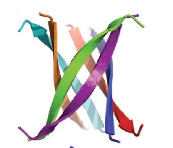
[[Image:Ribbonrepresentationofcrystalcylindrinfig2b.jpg | thumb | 400px | left | Fig. 3 Ribbon representation of the cylindrin crystal structure. ]] | [[Image:Ribbonrepresentationofcrystalcylindrinfig2b.jpg | thumb | 400px | left | Fig. 3 Ribbon representation of the cylindrin crystal structure. ]] | ||
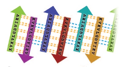
| - | [[Image:Unrolledcylindrin.jpg | thumb | | + | [[Image:Unrolledcylindrin.jpg | thumb | 500px | centre | Fig. 4 Schematic of unrolled cylindrin (outside view), illustrating strand-to-strand registration. Hydrogen bonds between the main chains of neighboring strands are shown by yellow dashed lines; hydrogen bonds mediated by water bridges or side chains are shown by blue dashed lines. ]] |
| + | {{ | ||
| + | |||
| + | }} | ||
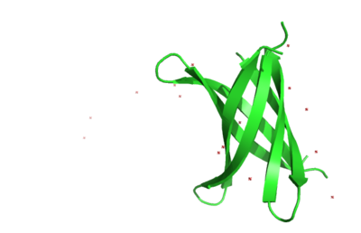
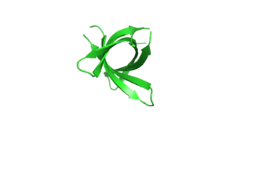
The structure of K11V-TR was determined, even though glycine linkers and Val-to-Leu replacement,cylindrical bodies of three double stranded K11V-TR oligomers and six-stranded K11V were identical. | The structure of K11V-TR was determined, even though glycine linkers and Val-to-Leu replacement,cylindrical bodies of three double stranded K11V-TR oligomers and six-stranded K11V were identical. | ||
| Line 40: | Line 43: | ||
[[Image:K11vtrsideview.png | thumb | 400px | centre | K11V-TR sideview ]] | [[Image:K11vtrsideview.png | thumb | 400px | centre | K11V-TR sideview ]] | ||
[[Image:Trtopview.png | thumb | 400px | centre | K11V-TR Top view ]] | [[Image:Trtopview.png | thumb | 400px | centre | K11V-TR Top view ]] | ||
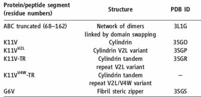
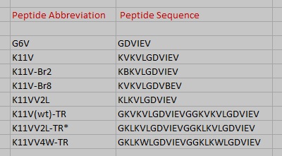
| - | [[Image:Names_all.jpg | thumb | | + | [[Image:Names_all.jpg | thumb | 400px | left | Fig. 2 : Cylindrin single chain and tandem repeat peptide abbreviations and amino acid sequences. ]] |
Revision as of 07:32, 15 November 2017
Toxic Amyloid Small Oligomer’s atomic view
| |||||||||||
References
- ↑ Laganowsky A, Liu C, Sawaya MR, Whitelegge JP, Park J, Zhao M, Pensalfini A, Soriaga AB, Landau M, Teng PK, Cascio D, Glabe C, Eisenberg D. Atomic view of a toxic amyloid small oligomer. Science. 2012 Mar 9;335(6073):1228-31. PMID:22403391 doi:10.1126/science.1213151
