Main Page
From Proteopedia
| Line 24: | Line 24: | ||
<tr style="font-size: 1.2em; text-align: center;"> | <tr style="font-size: 1.2em; text-align: center;"> | ||
| - | <td style="padding: 10px;background-color: #33ff7b"> | + | <td style="padding: 10px;background-color: #33ff7b"></td> |
| - | <td style="padding: 10px;background-color: #dae4d9"> | + | <td style="padding: 10px;background-color: #dae4d9"></td> |
| - | <td style="padding: 10px;background-color: #f1b840"> | + | <td style="padding: 10px;background-color: #f1b840"></td> |
| - | <td style="padding: 10px;background-color: #79baff"> | + | <td style="padding: 10px;background-color: #79baff"></td> |
</tr> | </tr> | ||
| Line 34: | Line 34: | ||
<td style="padding: 10px;> | <td style="padding: 10px;> | ||
| + | <p>[[:Category:Featured in Selected Pages|Other Selected Pages]]</p> | ||
<p>[[Help:Contents#For_authors:_contributing_content|How to author pages and contribute to Proteopedia]]</p> | <p>[[Help:Contents#For_authors:_contributing_content|How to author pages and contribute to Proteopedia]]</p> | ||
<p>[[Proteopedia:Video_Guide|Video Guides]]</p> | <p>[[Proteopedia:Video_Guide|Video Guides]]</p> | ||
| Line 40: | Line 41: | ||
<td style="padding: 10px;> | <td style="padding: 10px;> | ||
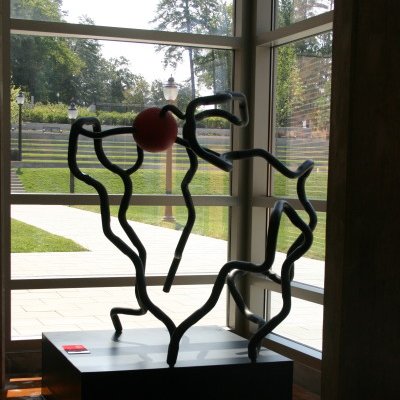
| + | <p>[[:Category:Featured in Art|Featured in Art]]</p> | ||
<p>[[:Category:PDB Art|All Art on Science]]</p> | <p>[[:Category:PDB Art|All Art on Science]]</p> | ||
</td> | </td> | ||
<td style="padding: 10px;> | <td style="padding: 10px;> | ||
| + | <p>[[:Category:Featured in I3DC|Featured in I3DC]]</p> | ||
<p>[[How to get an Interactive 3D Complement for your paper]]</p> | <p>[[How to get an Interactive 3D Complement for your paper]]</p> | ||
<p>[[Proteopedia:I3DC|List of Interactive Complements]]</p> | <p>[[Proteopedia:I3DC|List of Interactive Complements]]</p> | ||
| Line 50: | Line 53: | ||
<td style="padding: 10px;> | <td style="padding: 10px;> | ||
| + | <p>[[:Category:Featured in Education|Featured in Education]]</p> | ||
<p>[[Teaching Strategies Using Proteopedia]]</p> | <p>[[Teaching Strategies Using Proteopedia]]</p> | ||
<p>[[Teaching_Scenes%2C_Tutorials%2C_and_Educators%27_Pages|Examples of Pages for Teaching]]</p> | <p>[[Teaching_Scenes%2C_Tutorials%2C_and_Educators%27_Pages|Examples of Pages for Teaching]]</p> | ||
Revision as of 15:48, 18 October 2018
|
ISSN 2310-6301
Because life has more than 2D, Proteopedia helps to understand relationships between structure and function. Proteopedia is a free, collaborative 3D-encyclopedia of proteins & other molecules.
| |||||||||||
| Selected Pages | Art on Science | Journals | Education | ||||||||
|---|---|---|---|---|---|---|---|---|---|---|---|
|
|
|
|
||||||||
|
How to author pages and contribute to Proteopedia Who knows ... |
How to get an Interactive 3D Complement for your paper |
Teaching Strategies Using Proteopedia |
|||||||||
| |||||||||||