We apologize for Proteopedia being slow to respond. For the past two years, a new implementation of Proteopedia has been being built. Soon, it will replace this 18-year old system. All existing content will be moved to the new system at a date that will be announced here.
Sandbox Reserved 1613
From Proteopedia
(Difference between revisions)
| Line 15: | Line 15: | ||
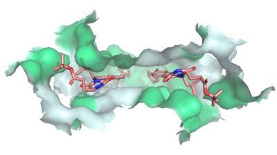
[[Image:Ligand_Interactions_6ffc.png|400 px|right|thumb|Figure 2: MZ29 bound to cavity 1 of ABCG2 (6ffc). Two MZ29 are shown in sticks and are colored by element. Hydrophobic interactions between the surface of cavity 1 and MZ29 are shown in green.]] | [[Image:Ligand_Interactions_6ffc.png|400 px|right|thumb|Figure 2: MZ29 bound to cavity 1 of ABCG2 (6ffc). Two MZ29 are shown in sticks and are colored by element. Hydrophobic interactions between the surface of cavity 1 and MZ29 are shown in green.]] | ||
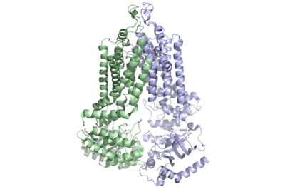
| - | Multidrug Transporter ABCG2 is a <scene name='83/832937/Dimer/1'>dimer</scene> that consists of two [https://en.wikipedia.org/wiki/Cavity cavities] separated by a <scene name='83/832937/Leucine_plug/4'>leucine plug</scene>. Cavity 1 is a binding pocket open to the [https://en.wikipedia.org/wiki/Cytoplasm cytoplasm] and the inner leaflet of the plasma membrane. Its shape is suitable to bind flat, hydrophobic and polycyclic substrates.<ref name="Manolaridis">PMID:30405239</ref> Many of its amino acids residues form hydrophobic interactions with the bound substrate, as shown in green in '''Figure 1'''. Cavity 2 is located above the leucine plug. It is empty until a <scene name='83/832937/Atp_and_mg_bound_to_abcg2/4'>magnesium ion and ATP</scene> are bound to ABCG2.<ref name=" Its <scene name='83/832937/Cysteine_disulfide_bridges/5'>inter- and intra-disulfides</scene> (yellow is inter- and intra-molecular disulfides, golden is intra-molecular only) promote the release of the substrate from the cavity into the extracellular space. | + | Multidrug Transporter ABCG2 is a <scene name='83/832937/Dimer/1'>dimer</scene> that consists of two [https://en.wikipedia.org/wiki/Cavity cavities] separated by a <scene name='83/832937/Leucine_plug/4'>leucine plug</scene>. Cavity 1 is a binding pocket open to the [https://en.wikipedia.org/wiki/Cytoplasm cytoplasm] and the inner leaflet of the plasma membrane. Its shape is suitable to bind flat, hydrophobic and polycyclic substrates.<ref name="Manolaridis">PMID:30405239</ref> Many of its amino acids residues form hydrophobic interactions with the bound substrate, as shown in green in '''Figure 1'''. Cavity 2 is located above the leucine plug. It is empty until a <scene name='83/832937/Atp_and_mg_bound_to_abcg2/4'>magnesium ion and ATP</scene> are bound to ABCG2.<ref name="Taylor">PMID:28554189</> Its <scene name='83/832937/Cysteine_disulfide_bridges/5'>inter- and intra-disulfides</scene> (yellow is inter- and intra-molecular disulfides, golden is intra-molecular only) promote the release of the substrate from the cavity into the extracellular space. |
One interesting feature of the NBD's is the fact that they remain in contact with one another even without a bound substrate. This makes the ABCG2 transporter unique and provides greater substrate specificity as the entrance to the transporter is not as globular as either ABCB1 or ABCC1. The entrance from the cytoplasm to the transporter is a [https://en.wikipedia.org/wiki/Hydrophobe hydrophobic] membrane entrance lined by <scene name='83/832939/Lining_of_entrance_of_nbd/1'>residues A397, V401, L405, L539, I543 and T547</scene> in both [https://en.wikipedia.org/wiki/Monomer monomers]. | One interesting feature of the NBD's is the fact that they remain in contact with one another even without a bound substrate. This makes the ABCG2 transporter unique and provides greater substrate specificity as the entrance to the transporter is not as globular as either ABCB1 or ABCC1. The entrance from the cytoplasm to the transporter is a [https://en.wikipedia.org/wiki/Hydrophobe hydrophobic] membrane entrance lined by <scene name='83/832939/Lining_of_entrance_of_nbd/1'>residues A397, V401, L405, L539, I543 and T547</scene> in both [https://en.wikipedia.org/wiki/Monomer monomers]. | ||
Dimerization of ABCG2 was originally thought to be achieved with the help of the <scene name='83/832939/Disproved_dimerization_process/1'>406xxx410 structural motif</scene> in each of the two domains but Cryo-EM showed that the [https://en.wikipedia.org/wiki/Sequence_motif motifs] were on opposite sides of the protein. | Dimerization of ABCG2 was originally thought to be achieved with the help of the <scene name='83/832939/Disproved_dimerization_process/1'>406xxx410 structural motif</scene> in each of the two domains but Cryo-EM showed that the [https://en.wikipedia.org/wiki/Sequence_motif motifs] were on opposite sides of the protein. | ||
Revision as of 13:33, 21 April 2020
ABCG2 Transporter Protein
| |||||||||||
References
- ↑ 1.0 1.1 1.2 Robey RW, Pluchino KM, Hall MD, Fojo AT, Bates SE, Gottesman MM. Revisiting the role of ABC transporters in multidrug-resistant cancer. Nat Rev Cancer. 2018 Jul;18(7):452-464. doi: 10.1038/s41568-018-0005-8. PMID:29643473 doi:http://dx.doi.org/10.1038/s41568-018-0005-8
- ↑ Manolaridis I, Jackson SM, Taylor NMI, Kowal J, Stahlberg H, Locher KP. Cryo-EM structures of a human ABCG2 mutant trapped in ATP-bound and substrate-bound states. Nature. 2018 Nov;563(7731):426-430. doi: 10.1038/s41586-018-0680-3. Epub 2018 Nov, 7. PMID:30405239 doi:http://dx.doi.org/10.1038/s41586-018-0680-3
- ↑ 3.0 3.1 PMID:28554189</> Its (yellow is inter- and intra-molecular disulfides, golden is intra-molecular only) promote the release of the substrate from the cavity into the extracellular space. One interesting feature of the NBD's is the fact that they remain in contact with one another even without a bound substrate. This makes the ABCG2 transporter unique and provides greater substrate specificity as the entrance to the transporter is not as globular as either ABCB1 or ABCC1. The entrance from the cytoplasm to the transporter is a hydrophobic membrane entrance lined by in both monomers. Dimerization of ABCG2 was originally thought to be achieved with the help of the in each of the two domains but Cryo-EM showed that the motifs were on opposite sides of the protein. <ref>PMID:30405239</li> <li id="cite_note-Jackson-3">↑ <sup>[[#cite_ref-Jackson_3-0|4.0]]</sup> <sup>[[#cite_ref-Jackson_3-1|4.1]]</sup> Jackson SM, Manolaridis I, Kowal J, Zechner M, Taylor NMI, Bause M, Bauer S, Bartholomaeus R, Bernhardt G, Koenig B, Buschauer A, Stahlberg H, Altmann KH, Locher KP. Structural basis of small-molecule inhibition of human multidrug transporter ABCG2. Nat Struct Mol Biol. 2018 Apr;25(4):333-340. doi: 10.1038/s41594-018-0049-1. Epub, 2018 Apr 2. PMID:[http://www.ncbi.nlm.nih.gov/pubmed/29610494 29610494] doi:[http://dx.doi.org/10.1038/s41594-018-0049-1 http://dx.doi.org/10.1038/s41594-018-0049-1]</li> <li id="cite_note-Fetsch-4">[[#cite_ref-Fetsch_4-0|↑]] Fetsch PA, Abati A, Litman T, Morisaki K, Honjo Y, Mittal K, Bates SE. Localization of the ABCG2 mitoxantrone resistance-associated protein in normal tissues. Cancer Lett. 2006 Apr 8;235(1):84-92. doi: 10.1016/j.canlet.2005.04.024. Epub, 2005 Jun 28. PMID:[http://www.ncbi.nlm.nih.gov/pubmed/15990223 15990223] doi:[http://dx.doi.org/10.1016/j.canlet.2005.04.024 http://dx.doi.org/10.1016/j.canlet.2005.04.024]</li> <li id="cite_note-Cleophas-5">[[#cite_ref-Cleophas_5-0|↑]] Cleophas MC, Joosten LA, Stamp LK, Dalbeth N, Woodward OM, Merriman TR. ABCG2 polymorphisms in gout: insights into disease susceptibility and treatment approaches. Pharmgenomics Pers Med. 2017 Apr 20;10:129-142. doi: 10.2147/PGPM.S105854., eCollection 2017. PMID:[http://www.ncbi.nlm.nih.gov/pubmed/28461764 28461764] doi:[http://dx.doi.org/10.2147/PGPM.S105854 http://dx.doi.org/10.2147/PGPM.S105854]</li></ol></ref>
Student Contributors
Shelby Skaggs, Samuel Sullivan, Jaelyn Voyles


