A Baculoviral System for the Production of Human β-Glucocerebrosidase Enables Atomic Resolution Analysis
Rhianna J. Rowland, Liang Wu, Feng Liu and Gideon J. Davies [1]
Molecular Tour
β-glucocerebrosidase (GBA) is a lysosomal enzyme which catalyses the hydrolysis of glycosphingolipids. Inherited deficiencies in GBA cause an accumulation of glycolipids in various cells throughout the body, subsequently leading to the most common lysosomal storage disorder Gaucher Disease (GD). Lysosomal storage disorders are a group of 50 rare inherited metabolic disorders which result from abnormal lysosomal function. GD affects multiple organs, such as the heart, kidneys, brain and liver, resulting in debilitating symptoms which severely impact an individuals quality of life and reduces their life expectancy.
Due to its involvement in GD, GBA is of considerable medical interest with continuous advances in the development of inhibitors, chaperones and activity-based probes for therapeutic and diagnostic applications. The development of novel GBA inhibitors requires a source of active protein, however, the majority of structural and mechanistic studies on this enzyme rely on expired clinical formulations which are incredibly costly and difficult to obtain in adequate supply. This problem is further compounded by a lack on consensus on a reliable platform for GBA production for research purposes.There is thus a need for an alternative source of GBA to meet research demands and reduce the reliance upon clinical formulations. As part of our long-standing interest in the development of activity-based probes to study GBA and other glycosidases, we established a baculovirus-insect cell expression system for the production of GBA.
The expression platform described herein produces active, crystallisable GBA which is comparable in enzymatic activity and biophysical properties to clinical preparations, rendering it. This protein crystallises readily and is amenable to ligand-binding studies, as demonstrated by a novel structure in complex with the glucosidase inhibitor, 2,4-dinitrophenyl-2-deoxy-2-fluoro-β-D-glucopyranoside. Furthermore, a novel crystal form of GBA which diffracts to give a 0.98 Å unliganded structure was described. This is the highest resolution structure of GBA deposited to date, permitting exquisite atomic resolution analysis which reveals two conformations of the catalytic acid-base residue. studies. In light of its purity, stability and activity, we envision that the baculoviral GBA production system described in this work will alleviate the over reliance on clinical formulations, aid in future biochemical studies of GBA and support structural-guided development of novel GBA ligands.
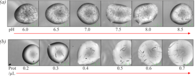
Figure 5 (a) Optimisation of crystallisation pH using bis-tris-propane buffer. (b) Optimisation of protein concentration.
(PDB 6tjk). N-glycans are shown in spacefill representation.
. Domain I (residues 1-27 and 383-414) in red, domain II (residues 30-75 and 431-497) in blue and domain III (residues 76-381 and 416-430) in gold. structure of bound bis-tris-propane which forms hydrogen bonds with Trp179, Asn234, Glu235, Glu340, Trp381 and an ethylene glycol (EDO) cryoprotectant molecules.
(PDB 6tjj).
(PDB 6tjq). The 2F-Glc moiety is covalently bound to the catalytic nucleophile (Glu340) which occupies two conformations. a/b = catalytic acid-base, Nuc = catalytic nucleophile, EDO = ethylene glycol.
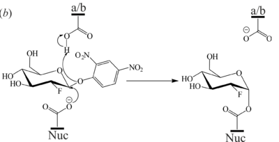
Figure 7 (b) Mechanism of 2F-DNPGlc hydrolysis by GBA to generate the covalent glycosyl-enzyme intermediate.
(PDB 6tn1). Domain I (residues 1-27 and 383-414) in orange, domain II (residues 30-75 and 431-497) in violet and domain III (residues 76–381 and 416-430) in blue. N-glycans are shown in ball-and-stick representation.
(PDB 6tjk).
References
- ↑ Rowland RJ, Wu L, Liu F, Davies GJ. A baculoviral system for the production of human beta-glucocerebrosidase enables atomic resolution analysis. Acta Crystallogr D Struct Biol. 2020 Jun 1;76(Pt 6):565-580. doi:, 10.1107/S205979832000501X. Epub 2020 May 29. PMID:32496218 doi:http://dx.doi.org/10.1107/S205979832000501X


