We apologize for Proteopedia being slow to respond. For the past two years, a new implementation of Proteopedia has been being built. Soon, it will replace this 18-year old system. All existing content will be moved to the new system at a date that will be announced here.
Sandbox Reserved 1621
From Proteopedia
(Difference between revisions)
| Line 16: | Line 16: | ||
===Substrate Structure=== | ===Substrate Structure=== | ||
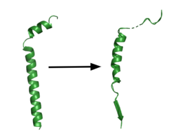
[[Image:App.png|250 px|right|thumb|'''Figure 1. APP fragment conformational change in gamma secretase.''' APP bound to GS undergoes a conformational change. The free state consists of 2 helices. Once bound to GS, the N-terminal helix unfolds into a coil and the C-terminal helix unwinds into a β-strand. Then the APP β-strand of APP forms a β-sheet interaction with PS1. Cleavage by the protease occurs between the helix and the β-strand.<ref name= "Zhou" /><ref name= "Nadezhdin" />]] | [[Image:App.png|250 px|right|thumb|'''Figure 1. APP fragment conformational change in gamma secretase.''' APP bound to GS undergoes a conformational change. The free state consists of 2 helices. Once bound to GS, the N-terminal helix unfolds into a coil and the C-terminal helix unwinds into a β-strand. Then the APP β-strand of APP forms a β-sheet interaction with PS1. Cleavage by the protease occurs between the helix and the β-strand.<ref name= "Zhou" /><ref name= "Nadezhdin" />]] | ||
| - | GS has been structurally characterized in the presence of both [https://en.wikipedia.org/wiki/Amyloid_precursor_protein APP] and Notch substrates.<ref name= "Zhou" /><ref name= "Yang" /> In each of these structures, the substrate bound in a similar location and underwent a similar structural transition upon binding to the active site of GS. Each substrate is composed of an N-terminal loop and a TM helix. The peptide substrate enters the enzyme by <scene name='83/832945/App_in_gs_general/3'>lateral diffusion</scene> via the lid complex, and once in place, the TM helix of the substrate is anchored by <scene name='83/832945/Hydrophobic_interactions/ | + | GS has been structurally characterized in the presence of both [https://en.wikipedia.org/wiki/Amyloid_precursor_protein APP] and Notch substrates.<ref name= "Zhou" /><ref name= "Yang" /> In each of these structures, the substrate bound in a similar location and underwent a similar structural transition upon binding to the active site of GS. Each substrate is composed of an N-terminal loop and a TM helix. The peptide substrate enters the enzyme by <scene name='83/832945/App_in_gs_general/3'>lateral diffusion</scene> via the lid complex, and once in place, the TM helix of the substrate is anchored by <scene name='83/832945/Hydrophobic_interactions/4'>van der Waals contacts</scene>. Upon binding to GS, the N-terminal extracellular helix of the substrate unwinds.<ref name="Nadezhdin">PMID:22649674</ref> The substrate's C-terminal end of the TM helix unwinds into a β-strand (Fig. 1). To differentiate substrates, the β-strand is often the main point of identification for the enzyme. Substrate binding induces a structural change in GS, creating two β-strands that form a β-sheet with the one β-strand of the substrate. This β-sheet is in close proximity with the active site, and guides the process of catalysis.<ref name="Zhou">PMID:30630874</ref> |
===Lid Complex=== | ===Lid Complex=== | ||
| Line 28: | Line 28: | ||
GS is connected with the development of AD in humans. Aβ fragment build up leads to [https://en.wikipedia.org/wiki/Amyloid amyloid]plaques in the brain.<ref name="Devendra">PMID:29477076</ref> Plaques in the brain cause severe neural dysfunction over time. | GS is connected with the development of AD in humans. Aβ fragment build up leads to [https://en.wikipedia.org/wiki/Amyloid amyloid]plaques in the brain.<ref name="Devendra">PMID:29477076</ref> Plaques in the brain cause severe neural dysfunction over time. | ||
| - | Mutations in GS are also connected with AD. Over 200 GS mutations have been linked to causing AD. These mutations target "hot spots" on the enzyme and seem to have a preponderance at the binding interface between <scene name='83/832945/Hydrophobic_interactions/ | + | Mutations in GS are also connected with AD. Over 200 GS mutations have been linked to causing AD. These mutations target "hot spots" on the enzyme and seem to have a preponderance at the binding interface between <scene name='83/832945/Hydrophobic_interactions/4'>PS1 and APP</scene>. The vast majority of these mutations are clustered in regions surrounding the C-terminal half of the <scene name='83/832945/Beta_sheet_complex/1'>APP TM helix and the β-strand</scene>. Mutations at these locations affect the integrity of APP recruitment and catalysis, implicating a role in the development of Aβ plaques that impair neural function.<ref name="Zhou" /> |
Inhibition of GS could be a potential AD treatment, but this would require targeting only APP cleavage over other GS substrates. APP cleavage leads to products such as Aβ42 and Aβ43,<ref name="Yang">PMID:28628788</ref> which are prone to aggregation and formation of Aβ plaques. Increased product peptide length contributes to aggregations, and many of the mutations within <scene name='83/832945/Ps1_subunit/1'>PS1</scene> result in elevated ratios of Aβ42 to the shorter Aβ40.<ref name="Bai">PMID:26280335</ref> The differential binding of APP and Notch to GS provides a starting point for differentiation but will require further follow-up studies to confirm that the structural differences observed are biologically relevant. Currently, to combat this complex situation, differences in binding between different substrates are being utilized to create drugs that selectively inhibit APP binding with GS, and possibly create a more ideal target for AD treatment.<ref name="Zhou">PMID:30630874</ref></StructureSection> | Inhibition of GS could be a potential AD treatment, but this would require targeting only APP cleavage over other GS substrates. APP cleavage leads to products such as Aβ42 and Aβ43,<ref name="Yang">PMID:28628788</ref> which are prone to aggregation and formation of Aβ plaques. Increased product peptide length contributes to aggregations, and many of the mutations within <scene name='83/832945/Ps1_subunit/1'>PS1</scene> result in elevated ratios of Aβ42 to the shorter Aβ40.<ref name="Bai">PMID:26280335</ref> The differential binding of APP and Notch to GS provides a starting point for differentiation but will require further follow-up studies to confirm that the structural differences observed are biologically relevant. Currently, to combat this complex situation, differences in binding between different substrates are being utilized to create drugs that selectively inhibit APP binding with GS, and possibly create a more ideal target for AD treatment.<ref name="Zhou">PMID:30630874</ref></StructureSection> | ||
Revision as of 23:53, 14 May 2020
Gamma Secretase
References
- ↑ 1.0 1.1 1.2 Bolduc DM, Montagna DR, Seghers MC, Wolfe MS, Selkoe DJ. The amyloid-beta forming tripeptide cleavage mechanism of gamma-secretase. Elife. 2016 Aug 31;5. doi: 10.7554/eLife.17578. PMID:27580372 doi:http://dx.doi.org/10.7554/eLife.17578
- ↑ 2.0 2.1 2.2 2.3 2.4 2.5 2.6 2.7 2.8 2.9 Zhou R, Yang G, Guo X, Zhou Q, Lei J, Shi Y. Recognition of the amyloid precursor protein by human gamma-secretase. Science. 2019 Feb 15;363(6428). pii: science.aaw0930. doi:, 10.1126/science.aaw0930. Epub 2019 Jan 10. PMID:30630874 doi:http://dx.doi.org/10.1126/science.aaw0930
- ↑ 3.0 3.1 Bai XC, Rajendra E, Yang G, Shi Y, Scheres SH. Sampling the conformational space of the catalytic subunit of human gamma-secretase. Elife. 2015 Dec 1;4. pii: e11182. doi: 10.7554/eLife.11182. PMID:26623517 doi:http://dx.doi.org/10.7554/eLife.11182
- ↑ 4.0 4.1 4.2 4.3 Bai XC, Yan C, Yang G, Lu P, Ma D, Sun L, Zhou R, Scheres SH, Shi Y. An atomic structure of human gamma-secretase. Nature. 2015 Aug 17. doi: 10.1038/nature14892. PMID:26280335 doi:http://dx.doi.org/10.1038/nature14892
- ↑ 5.0 5.1 5.2 5.3 5.4 Yang G, Zhou R, Zhou Q, Guo X, Yan C, Ke M, Lei J, Shi Y. Structural basis of Notch recognition by human gamma-secretase. Nature. 2019 Jan;565(7738):192-197. doi: 10.1038/s41586-018-0813-8. Epub 2018 Dec, 31. PMID:30598546 doi:http://dx.doi.org/10.1038/s41586-018-0813-8
- ↑ 6.0 6.1 Nadezhdin KD, Bocharova OV, Bocharov EV, Arseniev AS. Structural and dynamic study of the transmembrane domain of the amyloid precursor protein. Acta Naturae. 2011 Jan;3(1):69-76. PMID:22649674
- ↑ Kumar D, Ganeshpurkar A, Kumar D, Modi G, Gupta SK, Singh SK. Secretase inhibitors for the treatment of Alzheimer's disease: Long road ahead. Eur J Med Chem. 2018 Mar 25;148:436-452. doi: 10.1016/j.ejmech.2018.02.035. Epub , 2018 Feb 15. PMID:29477076 doi:http://dx.doi.org/10.1016/j.ejmech.2018.02.035
Student Contributors
Daniel Mulawa
Layla Wisser

