We apologize for Proteopedia being slow to respond. For the past two years, a new implementation of Proteopedia has been being built. Soon, it will replace this 18-year old system. All existing content will be moved to the new system at a date that will be announced here.
Sandbox Reserved 1700
From Proteopedia
(Difference between revisions)
| Line 19: | Line 19: | ||
=== Transmembrane Domain === | === Transmembrane Domain === | ||
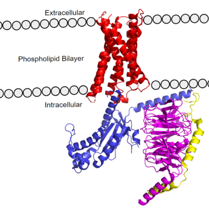
| - | The transmembrane domain spans the cell membrane ('''Figure 1''') and it consists of <scene name='90/904305/Transmembrane_protein_c_and_l/2'>seven transmembrane α-helices</scene> and <scene name='90/904305/Ecl_and_icl/3'>6 loops</scene> (three extracellular loops, and three intracellular loops). The transmembrane helices are numbered 1-7 and contain special conserved motifs that are shared across other A family receptors. These motifs are expanded upon later, as they heavily contribute to the structure and therefore function of the transmembrane domain as a whole. | + | The [https://en.wikipedia.org/wiki/Transmembrane_domain#:~:text=A%20transmembrane%20domain%20(TMD)%20is,can%20adopt%20a%20different%20conformation. transmembrane domain] spans the cell membrane ('''Figure 1''') and it consists of <scene name='90/904305/Transmembrane_protein_c_and_l/2'>seven transmembrane α-helices</scene> and <scene name='90/904305/Ecl_and_icl/3'>6 loops</scene> (three extracellular loops, and three intracellular loops). The transmembrane helices are numbered 1-7 and contain special conserved motifs that are shared across other A family receptors. These motifs are expanded upon later, as they heavily contribute to the structure and therefore function of the transmembrane domain as a whole. |
| - | The extracellular region of the 7 transmembrane domain forms a single binding pocket with <scene name='90/904305/Subpockets_1_and_2/4'>two sub-pockets</scene>. Sub-pocket 1 is negatively charged due to negatively charged <scene name='90/904305/Subpockets_1_and_2_d_and_e/2'>aspartate and glutamate</scene> residues (Asp-184 and Glu-164), while sub-pocket 2 contains hydrophobic amino acids which contribute to hydrophobic interactions between the ligand and protein. | + | The extracellular region of the 7 transmembrane domain forms a single [https://en.wikipedia.org/wiki/Binding_site binding pocket] with <scene name='90/904305/Subpockets_1_and_2/4'>two sub-pockets</scene>. Sub-pocket 1 is negatively charged due to negatively charged <scene name='90/904305/Subpockets_1_and_2_d_and_e/2'>aspartate and glutamate</scene> residues (Asp-184 and Glu-164), while sub-pocket 2 contains hydrophobic amino acids which contribute to hydrophobic interactions between the ligand and protein. |
The intracellular region ('''Figure 1''') is what connects the transmembrane helices with the G-protein. | The intracellular region ('''Figure 1''') is what connects the transmembrane helices with the G-protein. | ||
=== G-Protein === | === G-Protein === | ||
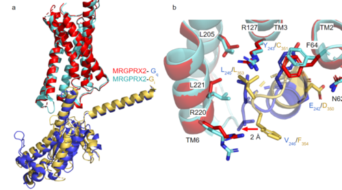
| - | [https://proteopedia.org/wiki/index.php/G_protein GTP-binding proteins], also known as G-proteins, are heterotrimeric complexes consisting of <scene name=''90/904305/Structure_overview_alpha/2'>alpha</scene>, <scene name='90/904305/Structure_overview_beta/2'>beta</scene>, and <scene name='90/904305/Structure_overview_gamma/2'>gamma</scene> subunits that interact with the intracellular transmembrane region at an <scene name='90/904306/Interface_2/1'>interface</scene> ( '''Figure 2b'''). G-proteins are responsible for transmitting extracellular signals into the cell upon activation. Activation leads to a substitution of GDP with GTP within the alpha subunit, causing the alpha subunit to disassociate from the beta and gamma subunits to initiate an intracellular signaling cascade. There are different families of G-alpha subunits, Gαi, Gαs, Gα12/13, and Gαq <ref name="Kamato">PMID: 26664886</ref>. MRGPRX2 binds to both Gαi and Gαq subunits with nearly identical structures despite slightly different amino acids present ( '''Figure 2a''') <ref name= "Cao" /> <ref name= "Yang" />. Throughout this page, MGPRX2 is always shown with Gq. The major difference between the Gq and Gi bound structures comes from one amino acid difference (valine on Gq versus phenylalanine on Gi) that pushes the Gi subunit 2Å away from the arginine residue on helix 6 of the transmembrane protein. | + | [https://proteopedia.org/wiki/index.php/G_protein GTP-binding proteins], also known as G-proteins, are heterotrimeric complexes consisting of <scene name=''90/904305/Structure_overview_alpha/2'>alpha</scene>, <scene name='90/904305/Structure_overview_beta/2'>beta</scene>, and <scene name='90/904305/Structure_overview_gamma/2'>gamma</scene> subunits that interact with the intracellular transmembrane region at an <scene name='90/904306/Interface_2/1'>interface</scene> ( '''Figure 2b'''). G-proteins are responsible for [https://en.wikipedia.org/wiki/Signal_transduction transmitting] extracellular signals into the cell upon activation. Activation leads to a substitution of GDP with GTP within the alpha subunit, causing the alpha subunit to disassociate from the beta and gamma subunits to initiate an intracellular signaling cascade. There are different families of G-alpha subunits, Gαi, Gαs, Gα12/13, and Gαq <ref name="Kamato">PMID: 26664886</ref>. MRGPRX2 binds to both Gαi and Gαq subunits with nearly identical structures despite slightly different amino acids present ( '''Figure 2a''') <ref name= "Cao" /> <ref name= "Yang" />. Throughout this page, MGPRX2 is always shown with Gq. The major difference between the Gq and Gi bound structures comes from one amino acid difference (valine on Gq versus phenylalanine on Gi) that pushes the Gi subunit 2Å away from the arginine residue on helix 6 of the transmembrane protein. |
| - | [[Image:Gq and gi overlay.png|500px|center|thumb|'''Figure 2a.''' Overlay of MGPRX2-Gq (red-dark blue) and MGPRX2-Gi (cyan-yellow). '''Figure 2b.''' Important residues involved in the interface between MGPRX2 and Gq/ Gi subunits. Arrow pointing to the major difference between the interfaces, which comes from the final C-terminus residue on the G-alpha subunit. In Gq, there is a valine while in Gi, there is a phenylalanine. This pushes the Gi subunit 2Å away from the arginine residue on helix 6 of the transmembrane protein.]] | + | [[Image:Gq and gi overlay.png|500px|center|thumb|'''Figure 2a.''' Overlay of MGPRX2-Gq (red-dark blue) and MGPRX2-Gi (cyan-yellow). '''Figure 2b.''' Important residues involved in the interface between MGPRX2 and Gq/ Gi subunits. Arrow pointing to the major difference between the interfaces, which comes from the final C-terminus residue on the G-alpha subunit. In Gq, there is a valine while in Gi, there is a phenylalanine. This pushes the Gi subunit 2Å away from the arginine residue on helix 6 of the transmembrane protein. All other interactions are nearly identical.]] |
===Class A Family Differences=== | ===Class A Family Differences=== | ||
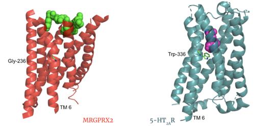
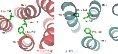
| - | The most important findings about the MRGPRX2 receptor are the differences between it and all other previously discovered GPCR's found in the A family. Many conserved structural motifs, characteristic of the A family receptors, are absent on MRGPRX2. These structural motif differences contribute to a membrane surface ligand binding rather than a ligand binding deep within the helices ('''Figure 3'''). To demonstrate this difference in depth binding, MRGPRX2 is compared to [https://proteopedia.org/wiki/index.php/Serotonin_receptor 5-HT2AR], another class A GPCR with more conserved structural motifs. | + | The most important findings about the MRGPRX2 receptor are the differences between it and all other previously discovered GPCR's found in the A family. Many conserved [https://en.wikipedia.org/wiki/Structural_motif structural motifs], characteristic of the A family receptors, are absent on MRGPRX2. These structural motif differences contribute to a membrane surface ligand binding rather than a ligand binding deep within the helices ('''Figure 3'''). To demonstrate this difference in depth binding, MRGPRX2 is compared to [https://proteopedia.org/wiki/index.php/Serotonin_receptor 5-HT2AR], another class A GPCR with more conserved structural motifs. |
---- | ---- | ||
---- | ---- | ||
Revision as of 20:12, 18 April 2022
MRGPRX2 Human Itch G-Protein Coupled Receptor (GPCR)
| |||||||||||
References
- ↑ Tuteja N. Signaling through G protein coupled receptors. Plant Signal Behav. 2009 Oct;4(10):942-7. doi: 10.4161/psb.4.10.9530. Epub 2009, Oct 14. PMID:19826234 doi:http://dx.doi.org/10.4161/psb.4.10.9530
- ↑ Hauser AS, Attwood MM, Rask-Andersen M, Schioth HB, Gloriam DE. Trends in GPCR drug discovery: new agents, targets and indications. Nat Rev Drug Discov. 2017 Dec;16(12):829-842. doi: 10.1038/nrd.2017.178. Epub, 2017 Oct 27. PMID:29075003 doi:http://dx.doi.org/10.1038/nrd.2017.178
- ↑ 3.0 3.1 3.2 Porebski G, Kwiecien K, Pawica M, Kwitniewski M. Mas-Related G Protein-Coupled Receptor-X2 (MRGPRX2) in Drug Hypersensitivity Reactions. Front Immunol. 2018 Dec 20;9:3027. doi: 10.3389/fimmu.2018.03027. eCollection, 2018. PMID:30619367 doi:http://dx.doi.org/10.3389/fimmu.2018.03027
- ↑ 4.0 4.1 4.2 4.3 4.4 4.5 4.6 Dondalska A, Ronnberg E, Ma H, Palsson SA, Magnusdottir E, Gao T, Adam L, Lerner EA, Nilsson G, Lagerstrom M, Spetz AL. Amelioration of Compound 48/80-Mediated Itch and LL-37-Induced Inflammation by a Single-Stranded Oligonucleotide. Front Immunol. 2020 Sep 30;11:559589. doi: 10.3389/fimmu.2020.559589. eCollection, 2020. PMID:33101278 doi:http://dx.doi.org/10.3389/fimmu.2020.559589
- ↑ 5.0 5.1 5.2 5.3 5.4 5.5 5.6 McNeil BD, Pundir P, Meeker S, Han L, Undem BJ, Kulka M, Dong X. Identification of a mast-cell-specific receptor crucial for pseudo-allergic drug reactions. Nature. 2015 Mar 12;519(7542):237-41. doi: 10.1038/nature14022. Epub 2014 Dec 17. PMID:25517090 doi:http://dx.doi.org/10.1038/nature14022
- ↑ 6.00 6.01 6.02 6.03 6.04 6.05 6.06 6.07 6.08 6.09 6.10 Cao C, Kang HJ, Singh I, Chen H, Zhang C, Ye W, Hayes BW, Liu J, Gumpper RH, Bender BJ, Slocum ST, Krumm BE, Lansu K, McCorvy JD, Kroeze WK, English JG, DiBerto JF, Olsen RHJ, Huang XP, Zhang S, Liu Y, Kim K, Karpiak J, Jan LY, Abraham SN, Jin J, Shoichet BK, Fay JF, Roth BL. Structure, function and pharmacology of human itch GPCRs. Nature. 2021 Dec;600(7887):170-175. doi: 10.1038/s41586-021-04126-6. Epub 2021, Nov 17. PMID:34789874 doi:http://dx.doi.org/10.1038/s41586-021-04126-6
- ↑ 7.0 7.1 7.2 7.3 7.4 7.5 7.6 7.7 7.8 Yang F, Guo L, Li Y, Wang G, Wang J, Zhang C, Fang GX, Chen X, Liu L, Yan X, Liu Q, Qu C, Xu Y, Xiao P, Zhu Z, Li Z, Zhou J, Yu X, Gao N, Sun JP. Structure, function and pharmacology of human itch receptor complexes. Nature. 2021 Dec;600(7887):164-169. doi: 10.1038/s41586-021-04077-y. Epub 2021, Nov 17. PMID:34789875 doi:http://dx.doi.org/10.1038/s41586-021-04077-y
- ↑ Kamato D, Thach L, Bernard R, Chan V, Zheng W, Kaur H, Brimble M, Osman N, Little PJ. Structure, Function, Pharmacology, and Therapeutic Potential of the G Protein, Galpha/q,11. Front Cardiovasc Med. 2015 Mar 24;2:14. doi: 10.3389/fcvm.2015.00014. eCollection, 2015. PMID:26664886 doi:http://dx.doi.org/10.3389/fcvm.2015.00014
- ↑ Trzaskowski B, Latek D, Yuan S, Ghoshdastider U, Debinski A, Filipek S. Action of molecular switches in GPCRs--theoretical and experimental studies. Curr Med Chem. 2012;19(8):1090-109. doi: 10.2174/092986712799320556. PMID:22300046 doi:http://dx.doi.org/10.2174/092986712799320556
- ↑ Olivella M, Caltabiano G, Cordomi A. The role of Cysteine 6.47 in class A GPCRs. BMC Struct Biol. 2013 Mar 15;13:3. doi: 10.1186/1472-6807-13-3. PMID:23497259 doi:http://dx.doi.org/10.1186/1472-6807-13-3
- ↑ Gonzalez-Rey E, Chorny A, Robledo G, Delgado M. Cortistatin, a new antiinflammatory peptide with therapeutic effect on lethal endotoxemia. J Exp Med. 2006 Mar 20;203(3):563-71. doi: 10.1084/jem.20052017. Epub 2006 Feb, 21. PMID:16492802 doi:http://dx.doi.org/10.1084/jem.20052017
