User:Glauco O. Gavioli Ferreira/Sandbox 1
From Proteopedia
(Difference between revisions)
| Line 15: | Line 15: | ||
[[Image:Alinhamento focoGhelice.png|thumb|right|554x320px|<b>Fig. 1</b> - Maspin on its closed (PDB code [[1xqg]]) and opened (PDB code [[1wz9]]) form based on the ideia of the conformation "switch" in wich the rotation of G-helix changes maspin's charge distribuition <ref name="P" />. <font color='blue'><b>Positive</b></font> and <font color='red'><b>negative</b></font> charges can be seen in blue and red respectively, likewise <span style="color:white;background-color:darkgrey;font-weight:bold;">neutral net charge</span> are represented by whyte color. The region comprising the G-helix is <span style="color:black;background-color:yellow;font-weight:bold;">highlighted</span>.]] | [[Image:Alinhamento focoGhelice.png|thumb|right|554x320px|<b>Fig. 1</b> - Maspin on its closed (PDB code [[1xqg]]) and opened (PDB code [[1wz9]]) form based on the ideia of the conformation "switch" in wich the rotation of G-helix changes maspin's charge distribuition <ref name="P" />. <font color='blue'><b>Positive</b></font> and <font color='red'><b>negative</b></font> charges can be seen in blue and red respectively, likewise <span style="color:white;background-color:darkgrey;font-weight:bold;">neutral net charge</span> are represented by whyte color. The region comprising the G-helix is <span style="color:black;background-color:yellow;font-weight:bold;">highlighted</span>.]] | ||
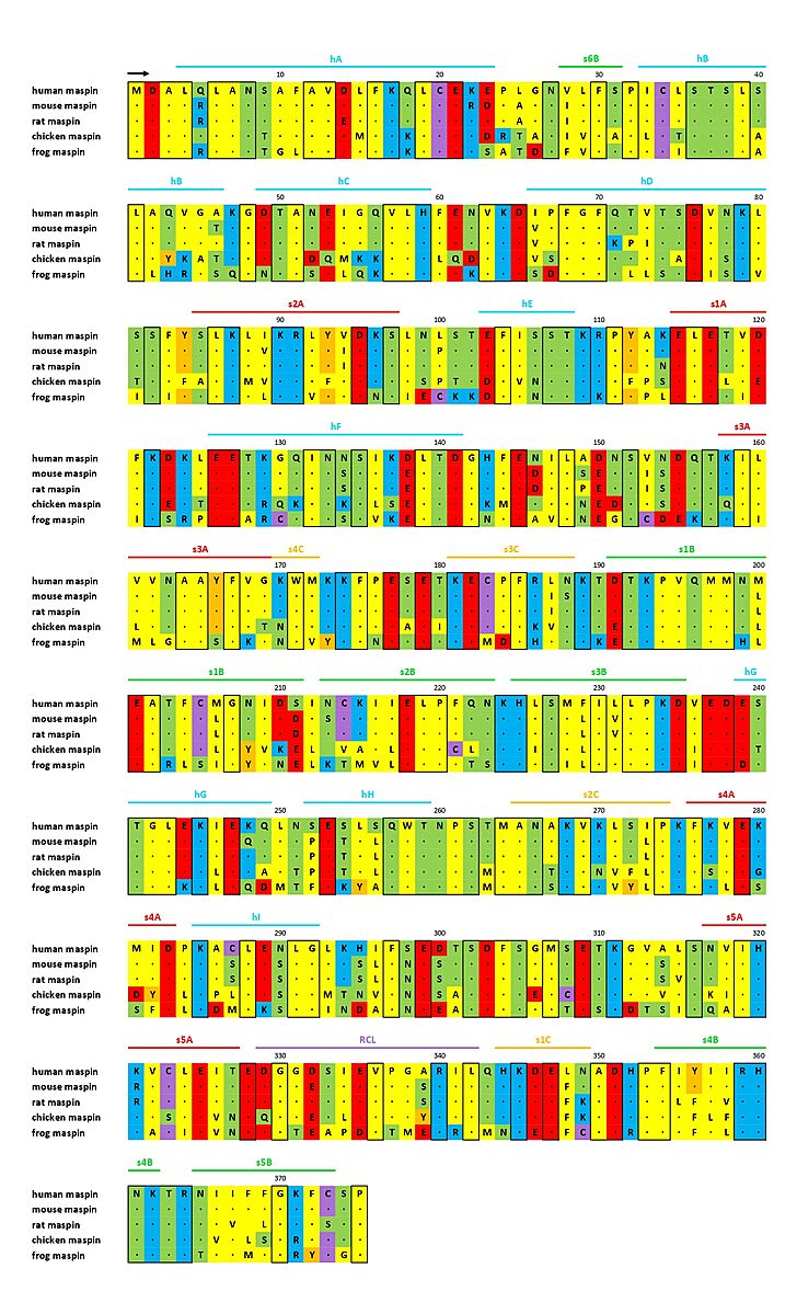
| - | Also maspin is not limited to a certain cell compartment, once it is found on nucleus, cytoplasm, membrane, and as a secreted protein, according to the cell type and tissue <ref name="numero1" /><ref name="numero2" />. Currently, it is known that the subcellular location of maspin is important for its <b>tumor suppressor activity</b>, and not only its protein levels inside the cell. In the past, there was a controversy about it, once maspin was upregulated in some tumors, while downregulated in others. Then, its translocation to the nucleus was observed and maspin’s nuclear localization was related to its tumor suppressor function <ref name="numero8">PMID: 21502940</ref>. However, contrary to what is expected, it has never been found a nuclear localization sequence (NLS), nuclear export sequence (NES), neither a secretory leader sequence (SLS) on maspin structure <ref name="numero4">PMID: 22752408</ref>. A map of maspin-like proteins is shown in Figure | + | Also maspin is not limited to a certain cell compartment, once it is found on nucleus, cytoplasm, membrane, and as a secreted protein, according to the cell type and tissue <ref name="numero1" /><ref name="numero2" />. Currently, it is known that the subcellular location of maspin is important for its <b>tumor suppressor activity</b>, and not only its protein levels inside the cell. In the past, there was a controversy about it, once maspin was upregulated in some tumors, while downregulated in others. Then, its translocation to the nucleus was observed and maspin’s nuclear localization was related to its tumor suppressor function <ref name="numero8">PMID: 21502940</ref>. However, contrary to what is expected, it has never been found a nuclear localization sequence (NLS), nuclear export sequence (NES), neither a secretory leader sequence (SLS) on maspin structure <ref name="numero4">PMID: 22752408</ref>. A map of maspin-like proteins is shown in Figure 2. |
The tumor suppressor function of maspin is probably related to its activities, which are mainly inhibition of cell growth, invasion, tumoral migration, apoptosis stimuli, gene transcription regulation, angiogenesis inhibition <ref name="numero4" /> and prevention of oxidative damage of the proteome <ref name="numero5">PMID: 16049007</ref>. Besides all of these functions, maspin also has an important role in the organization of the epiblast during early embryonic development. However, maspin lacks studies on non-tumoral cell lines, and its role on a normal condition might be different from its activity inside a tumoral lineages <ref name="P" />. | The tumor suppressor function of maspin is probably related to its activities, which are mainly inhibition of cell growth, invasion, tumoral migration, apoptosis stimuli, gene transcription regulation, angiogenesis inhibition <ref name="numero4" /> and prevention of oxidative damage of the proteome <ref name="numero5">PMID: 16049007</ref>. Besides all of these functions, maspin also has an important role in the organization of the epiblast during early embryonic development. However, maspin lacks studies on non-tumoral cell lines, and its role on a normal condition might be different from its activity inside a tumoral lineages <ref name="P" />. | ||
| - | [[Image:Imagem1_mapa.jpg|thumb|center|729x1200px|<b>Fig. | + | [[Image:Imagem1_mapa.jpg|thumb|center|729x1200px|<b>Fig. 2</b> - Sequence alignment of maspin-like proteins. The secondary structure of maspin is marked above the alignment and conserved residues are enclosed in a black box (ADAPTED FROM doi: http://dx.doi.org/10.1074/jbc.M412043200).]] |
| Line 47: | Line 47: | ||
| - | [[Image:Imagem_Ghelice_aberta_vs_fechada1.png|thumb|left|578x325px|<b>Fig. | + | [[Image:Imagem_Ghelice_aberta_vs_fechada1.png|thumb|left|578x325px|<b>Fig. 3</b> - Maspin on its closed (PDB code [[1xqg]]) and opened (PDB code [[1wz9]]) form based on the ideia of the conformation "switch" in wich the rotation of G-helix changes maspin's charge distribuition <ref name="P" />. <font color='blue'><b>Positive</b></font> and <font color='red'><b>negative</b></font> charges can be seen in blue and red respectively, likewise <span style="color:white;background-color:darkgrey;font-weight:bold;">neutral net charge</span> are represented by whyte color. The region comprising the G-helix is <span style="color:black;background-color:yellow;font-weight:bold;">highlighted</span>.]] |
====G-helix==== | ====G-helix==== | ||
Maspin is able to undergo conformational change in and around the <scene name='91/915218/G_helix/2'>G-helix</scene>, presenting two shapes: <b>an <scene name='91/915218/Open_masp_cartoon/3'>opened</scene> and a <scene name='91/915218/Closed_masp_cartoon/3'>closed</scene> form</b>. This is a real putative cofactor binding site and may determine maspin’s function. | Maspin is able to undergo conformational change in and around the <scene name='91/915218/G_helix/2'>G-helix</scene>, presenting two shapes: <b>an <scene name='91/915218/Open_masp_cartoon/3'>opened</scene> and a <scene name='91/915218/Closed_masp_cartoon/3'>closed</scene> form</b>. This is a real putative cofactor binding site and may determine maspin’s function. | ||
| - | Once the region at and around the G-helix has flexibility to undergo a conformational change, as a consequence, its <scene name='91/915218/G_helix_charged/1'>charged residues</scene> (where <font color='blue'><b>positive</b></font> and <font color='red'><b>negative</b></font> charges, as well as <span style="color:white;background-color:darkgrey;font-weight:bold;">neutral net charge</span> are highlighted) are reorganized and the central part of the helix structure becomes negative. The two structures, reflecting maspin in the open and closed conformation, show that rotation of the G-helix alters the local charge distribution (Fig. | + | Once the region at and around the G-helix has flexibility to undergo a conformational change, as a consequence, its <scene name='91/915218/G_helix_charged/1'>charged residues</scene> (where <font color='blue'><b>positive</b></font> and <font color='red'><b>negative</b></font> charges, as well as <span style="color:white;background-color:darkgrey;font-weight:bold;">neutral net charge</span> are highlighted) are reorganized and the central part of the helix structure becomes negative. The two structures, reflecting maspin in the open and closed conformation, show that rotation of the G-helix alters the local charge distribution (Fig. 3), suggesting that this movement represents a conformational “switch” (as we can see on these spacefill representations of <scene name='91/915218/Open_maspin/2'>opened</scene> and <scene name='91/915218/Closed_maspin/2'>closed</scene> forms of maspin), what researchers have implied be a cofactor binding site under conformational control based on G-helix negative charged patch modulation <ref name="P" />. |
Revision as of 06:20, 15 July 2022
SerpinB5 (Maspin)
| |||||||||||
References
- ↑ 1.0 1.1 1.2 Khalkhali-Ellis Z. Maspin: the new frontier. Clin Cancer Res. 2006 Dec 15;12(24):7279-83. doi: 10.1158/1078-0432.CCR-06-1589. PMID:17189399 doi:http://dx.doi.org/10.1158/1078-0432.CCR-06-1589
- ↑ 2.0 2.1 Banias L, Jung I, Gurzu S. Subcellular expression of maspin – from normal tissue to tumor cells. World J Meta-Anal 2019; 7(4): 142-155. doi: https://dx.doi.org/10.13105/wjma.v7.i4.142
- ↑ 3.0 3.1 3.2 3.3 3.4 3.5 3.6 3.7 3.8 3.9 Law RH, Irving JA, Buckle AM, Ruzyla K, Buzza M, Bashtannyk-Puhalovich TA, Beddoe TC, Nguyen K, Worrall DM, Bottomley SP, Bird PI, Rossjohn J, Whisstock JC. The high resolution crystal structure of the human tumor suppressor maspin reveals a novel conformational switch in the G-helix. J Biol Chem. 2005 Jun 10;280(23):22356-64. Epub 2005 Mar 10. PMID:15760906 doi:http://dx.doi.org/10.1074/jbc.M412043200
- ↑ 4.0 4.1 4.2 4.3 Law RH, Zhang Q, McGowan S, Buckle AM, Silverman GA, Wong W, Rosado CJ, Langendorf CG, Pike RN, Bird PI, Whisstock JC. An overview of the serpin superfamily. Genome Biol. 2006;7(5):216. doi: 10.1186/gb-2006-7-5-216. Epub 2006 May 30. PMID:16737556 doi:http://dx.doi.org/10.1186/gb-2006-7-5-216
- ↑ Huntington JA, Read RJ, Carrell RW. Structure of a serpin-protease complex shows inhibition by deformation. Nature. 2000 Oct 19;407(6806):923-6. PMID:11057674 doi:10.1038/35038119
- ↑ Zou Z, Anisowicz A, Hendrix MJ, Thor A, Neveu M, Sheng S, Rafidi K, Seftor E, Sager R. Maspin, a serpin with tumor-suppressing activity in human mammary epithelial cells. Science. 1994 Jan 28;263(5146):526-9. PMID:8290962
- ↑ Goulet B, Kennette W, Ablack A, Postenka CO, Hague MN, Mymryk JS, Tuck AB, Giguere V, Chambers AF, Lewis JD. Nuclear localization of maspin is essential for its inhibition of tumor growth and metastasis. Lab Invest. 2011 Aug;91(8):1181-7. doi: 10.1038/labinvest.2011.66. Epub 2011 Apr , 18. PMID:21502940 doi:http://dx.doi.org/10.1038/labinvest.2011.66
- ↑ 8.0 8.1 Bodenstine TM, Seftor RE, Khalkhali-Ellis Z, Seftor EA, Pemberton PA, Hendrix MJ. Maspin: molecular mechanisms and therapeutic implications. Cancer Metastasis Rev. 2012 Dec;31(3-4):529-51. doi: 10.1007/s10555-012-9361-0. PMID:22752408 doi:http://dx.doi.org/10.1007/s10555-012-9361-0
- ↑ 9.0 9.1 9.2 Yin S, Li X, Meng Y, Finley RL Jr, Sakr W, Yang H, Reddy N, Sheng S. Tumor-suppressive maspin regulates cell response to oxidative stress by direct interaction with glutathione S-transferase. J Biol Chem. 2005 Oct 14;280(41):34985-96. doi: 10.1074/jbc.M503522200. Epub 2005, Jul 26. PMID:16049007 doi:http://dx.doi.org/10.1074/jbc.M503522200
- ↑ Longhi MT, Silva LE, Pereira M, Magalhaes M, Reina J, Vitorino FNL, Gumbiner BM, da Cunha JPC, Cella N. PI3K-AKT, JAK2-STAT3 pathways and cell-cell contact regulate maspin subcellular localization. Cell Commun Signal. 2021 Aug 14;19(1):86. doi: 10.1186/s12964-021-00758-3. PMID:34391444 doi:http://dx.doi.org/10.1186/s12964-021-00758-3
- ↑ Al-Ayyoubi M, Gettins PG, Volz K. Crystal structure of human maspin, a serpin with antitumor properties: reactive center loop of maspin is exposed but constrained. J Biol Chem. 2004 Dec 31;279(53):55540-4. Epub 2004 Oct 22. PMID:15501821 doi:10.1074/jbc.M409957200
- ↑ Silverman GA, Bird PI, Carrell RW, Church FC, Coughlin PB, Gettins PG, Irving JA, Lomas DA, Luke CJ, Moyer RW, Pemberton PA, Remold-O'Donnell E, Salvesen GS, Travis J, Whisstock JC. The serpins are an expanding superfamily of structurally similar but functionally diverse proteins. Evolution, mechanism of inhibition, novel functions, and a revised nomenclature. J Biol Chem. 2001 Sep 7;276(36):33293-6. doi: 10.1074/jbc.R100016200. Epub 2001, Jul 2. PMID:11435447 doi:http://dx.doi.org/10.1074/jbc.R100016200
- ↑ Sheng S, Pemberton PA, Sager R. Production, purification, and characterization of recombinant maspin proteins. J Biol Chem. 1994 Dec 9;269(49):30988-93. PMID:7983035
- ↑ Pemberton PA, Wong DT, Gibson HL, Kiefer MC, Fitzpatrick PA, Sager R, Barr PJ. The tumor suppressor maspin does not undergo the stressed to relaxed transition or inhibit trypsin-like serine proteases. Evidence that maspin is not a protease inhibitory serpin. J Biol Chem. 1995 Jun 30;270(26):15832-7. PMID:7797587
- ↑ Bass R, Fernandez AM, Ellis V. Maspin inhibits cell migration in the absence of protease inhibitory activity. J Biol Chem. 2002 Dec 6;277(49):46845-8. doi: 10.1074/jbc.C200532200. Epub 2002, Oct 15. PMID:12384513 doi:http://dx.doi.org/10.1074/jbc.C200532200
- ↑ 16.0 16.1 Ngamkitidechakul C, Warejcka DJ, Burke JM, O'Brien WJ, Twining SS. Sufficiency of the reactive site loop of maspin for induction of cell-matrix adhesion and inhibition of cell invasion. Conversion of ovalbumin to a maspin-like molecule. J Biol Chem. 2003 Aug 22;278(34):31796-806. doi: 10.1074/jbc.M302408200. Epub, 2003 Jun 10. PMID:12799381 doi:http://dx.doi.org/10.1074/jbc.M302408200
- ↑ 17.0 17.1 17.2 Al-Ayyoubi M, Gettins PG, Volz K. Crystal structure of human maspin, a serpin with antitumor properties: reactive center loop of maspin is exposed but constrained. J Biol Chem. 2004 Dec 31;279(53):55540-4. Epub 2004 Oct 22. PMID:15501821 doi:10.1074/jbc.M409957200
- ↑ 18.0 18.1 Dzinic SH, Kaplun A, Li X, Bernardo M, Meng Y, Dean I, Krass D, Stemmer P, Shin N, Lonardo F, Sheng S. Identification of an intrinsic determinant critical for maspin subcellular localization and function. PLoS One. 2013 Nov 21;8(11):e74502. doi: 10.1371/journal.pone.0074502., eCollection 2013. PMID:24278104 doi:http://dx.doi.org/10.1371/journal.pone.0074502
- ↑ Li X, Yin S, Meng Y, Sakr W, Sheng S. Endogenous inhibition of histone deacetylase 1 by tumor-suppressive maspin. Cancer Res. 2006 Sep 15;66(18):9323-9. doi: 10.1158/0008-5472.CAN-06-1578. PMID:16982778 doi:http://dx.doi.org/10.1158/0008-5472.CAN-06-1578
- ↑ Bernardo MM, Meng Y, Lockett J, Dyson G, Dombkowski A, Kaplun A, Li X, Yin S, Dzinic S, Olive M, Dean I, Krass D, Moin K, Bonfil RD, Cher M, Sakr W, Sheng S. Maspin reprograms the gene expression profile of prostate carcinoma cells for differentiation. Genes Cancer. 2011 Nov;2(11):1009-22. doi: 10.1177/1947601912440170. PMID:22737267 doi:http://dx.doi.org/10.1177/1947601912440170
- ↑ Lee SJ, Jang H, Park C. Maspin increases Ku70 acetylation and Bax-mediated cell death in cancer cells. Int J Mol Med. 2012 Feb;29(2):225-30. doi: 10.3892/ijmm.2011.833. Epub 2011 Nov, 10. PMID:22076034 doi:http://dx.doi.org/10.3892/ijmm.2011.833
- ↑ Singhal SS, Singh SP, Singhal P, Horne D, Singhal J, Awasthi S. Antioxidant role of glutathione S-transferases: 4-Hydroxynonenal, a key molecule in stress-mediated signaling. Toxicol Appl Pharmacol. 2015 Dec 15;289(3):361-70. doi:, 10.1016/j.taap.2015.10.006. Epub 2015 Oct 23. PMID:26476300 doi:http://dx.doi.org/10.1016/j.taap.2015.10.006
- ↑ Nawata S, Shi HY, Sugino N, Zhang M. Evidence of post-translational modification of the tumor suppressor maspin under oxidative stress. Int J Mol Med. 2011 Feb;27(2):249-54. doi: 10.3892/ijmm.2010.572. Epub 2010 Dec, 2. PMID:21132260 doi:http://dx.doi.org/10.3892/ijmm.2010.572


