This old version of Proteopedia is provided for student assignments while the new version is undergoing repairs. Content and edits done in this old version of Proteopedia after March 1, 2026 will eventually be lost when it is retired in about June of 2026.
Apply for new accounts at the new Proteopedia. Your logins will work in both the old and new versions.
Green Fluorescent Protein
From Proteopedia
| Line 80: | Line 80: | ||
Green fluorescent protein (<scene name='Green_Fluorescent_Protein/Initial/1'>default scene</scene>) is a 21 kDa protein consisting of 238 residues strung together to form a | Green fluorescent protein (<scene name='Green_Fluorescent_Protein/Initial/1'>default scene</scene>) is a 21 kDa protein consisting of 238 residues strung together to form a | ||
<scene name='Green_Fluorescent_Protein/Secondary_structure/1'>secondary structure</scene> of five α-helices and one eleven-stranded β-pleated sheet,<ref name="PDBsum" /> where each strand contains nine to thirteen residues each.<ref name="Ormo" /> (To view the primary and secondary structure of GFP, go to [[http://www.ebi.ac.uk/thornton-srv/databases/cgi-bin/pdbsum/GetPage.pl?pdbcode=1ema&template=protein.html&r=wiring&l=1&chain=A www.ebi.aci.uk]].) These β-strands display an almost “seamless symmetry” in which only two of the strands vary in structural content.<ref name="Phillips">Phillips GN Jr. 1997. Structure and dynamics of green fluorescent protein. Curr Opin Struct Biol. 7(6):821-827. DOI 10.1016/S0959-440X(97)80153-4.</ref> This β-sheet conforms itself through regular hydrogen bonding into a β-barrel.<ref name="Yang" /> In GFP, the structure is so regular that <scene name='Green_Fluorescent_Protein/Water_stripes/1'>"stripes"</scene> of water molecules (red) can be seen following the structure of the barrel.<ref name="Phillips" /> Together with the α-helices at either end of the molecule, a nearly perfect cylinder is produced, 42Å long and 24Å in diameter,<ref name="Ormo" /> creating what is referred to as a “β-can” formation.<ref name="Phillips" /> The short helical segments at either end of the cylinder form “caps” to further protect the interior of the β-barrel.<ref name="Phillips" /> Overall stability is maintained by this β-can structure, helping to resist unfolding from heat and other denaturants.<ref name="Yang" /> | <scene name='Green_Fluorescent_Protein/Secondary_structure/1'>secondary structure</scene> of five α-helices and one eleven-stranded β-pleated sheet,<ref name="PDBsum" /> where each strand contains nine to thirteen residues each.<ref name="Ormo" /> (To view the primary and secondary structure of GFP, go to [[http://www.ebi.ac.uk/thornton-srv/databases/cgi-bin/pdbsum/GetPage.pl?pdbcode=1ema&template=protein.html&r=wiring&l=1&chain=A www.ebi.aci.uk]].) These β-strands display an almost “seamless symmetry” in which only two of the strands vary in structural content.<ref name="Phillips">Phillips GN Jr. 1997. Structure and dynamics of green fluorescent protein. Curr Opin Struct Biol. 7(6):821-827. DOI 10.1016/S0959-440X(97)80153-4.</ref> This β-sheet conforms itself through regular hydrogen bonding into a β-barrel.<ref name="Yang" /> In GFP, the structure is so regular that <scene name='Green_Fluorescent_Protein/Water_stripes/1'>"stripes"</scene> of water molecules (red) can be seen following the structure of the barrel.<ref name="Phillips" /> Together with the α-helices at either end of the molecule, a nearly perfect cylinder is produced, 42Å long and 24Å in diameter,<ref name="Ormo" /> creating what is referred to as a “β-can” formation.<ref name="Phillips" /> The short helical segments at either end of the cylinder form “caps” to further protect the interior of the β-barrel.<ref name="Phillips" /> Overall stability is maintained by this β-can structure, helping to resist unfolding from heat and other denaturants.<ref name="Yang" /> | ||
| + | |||
| + | One <scene name='Green_Fluorescent_Protein/Central_helix/1'>α-helix</scene> can be found running through the central axis of the β-barrel,<ref name="Haldar" /> roughly <scene name='Green_Fluorescent_Protein/Perpendicular/1'>perpendicular</scene> to the symmetry axis of the barrel.<ref name="Ormo">Ormo M, Cubitt AB, Kallio K, Gross LA, Tsien RY, Remington SJ. 1996. Crystal structure of the ''Aequorea victoria'' green fluorescent protein. Science. 273(5280):1392-1395. DOI 10.1126/science.273.5280.1392.</ref> This helix is extremely important as it contains the fluorophore responsible for fluorescence.<ref name="Yang" /><ref name="Haldar" /> This α-helix in particular is highly stabilized by the many <scene name='Green_Fluorescent_Protein/Spacefill/1'>contacts</scene> that are made with each strand of the barrel.<ref name="Andrews">Andrews BT, Gosavi S, Finke JM, Onuchic JN, Jennings PA. 2008. The dual-basin landscape in GFP. Proceedings of the National Academy of Sciences. 105(34):12283-12288. DOI 10.1073/pnas.0804039105.</ref> | ||
| + | |||
| + | ===The Chromophore=== | ||
| + | |||
| + | The <scene name='Green_Fluorescent_Protein/Chromophore/1'>chromophore</scene> of GFP is located at the center of the β-barrel with a wild-type excitation peak of 395 nm, and a minor peak at 475 nm (about three times less intense<ref name="Tsien" />) <ref name="Yang" /><ref name="Cubitt" /><ref name="Ormo" /><ref name="Phillips" /> with extinction coefficients of approximately 30,000 and 7,000 M<sup>-1</sup> cm<sup>-1</sup>, respectively.<ref name="Yang" /><ref name="Phillips" /> Interestingly, the ''Aequorea victoria'' jellyfish utilizes the smaller of the two excitation peaks as pure aequorin emits a light of 470 nm.<ref name="Tsien">Tsien, Roger Y. 1998. The Green Fluorescent Protein. Annual Review in Biochemistry. 67:509-544.</ref> The relative amplitudes of these two excitation peaks can vary depending on environmental factors and previous illumination.<ref name="Ormo" /> For example, continued excitation leads to a diminution of the 395 nm excitation peak with a reciprocal amplification of the 475 nm peak.<ref name="Phillips" /> Regardless of absorption, the chromophore of GFP emits light of 508 nm.<ref name="Yang" /><ref name="Cubitt" /><ref name="Ormo" /><ref name="Phillips" /> | ||
| + | |||
| + | Three amino residues in the central α-helix constitute the fluorophore of GFP: Ser<sup>65</sup>Tyr<sup>66</sup>Gly<sup>67</sup> (see left). Tsien et al. discovered that this tri-peptide sequence is post-translationally modified by internal cyclization and oxidation<ref name="Haldar" /> to produce a <scene name='Green_Fluorescent_Protein/Chromophore_structure/1'>4-(p-hydroxybenzylidene)-imidazolidin-5-one</scene> structure.<ref name="Yang" /> Studies with E. coli proposed a sequential mechanism for the formation of the fluorophore that was initiated by a rapid cyclization between Ser<sup>65</sup> and Gly<sup>67</sup> to form an imidazolin-5-one intermediate.<ref name="Yang" /> This rapid cyclization is carried out via nucleophilic attack of the amino group from Gly<sup>67</sup> on the carbonyl group of Ser<sup>65</sup> to form a five-membered ring. The loss of water then forms the imidazolin-5-one intermediate.<ref name="Cubitt" /> Cyclization is succeeded by a much slower rate-limiting oxygenation of the Tyr<sup>66</sup> hydroxybenzyl side chain by atmospheric oxygen (No fluorescence was seen in anaerobically grown E. coli.), resulting in the 4-(p-hydroxybenzylidene)-imidazolidin-5-one stucture.<ref name="Yang" /><ref name="Cubitt" /><ref name="Phillips" /> The double bond that results from this series of reactions results in the linkage of the two π-systems of the rings, forming a larger conjugated system essential for fluorophore stability. <ref name="Bublitz"> Bublitz G, King BA, Boxer SG. 1998. Electronic structure of the chromophore in green fluorescent protein (GFP). Journal of the American Chemical Society. 120(36): 9370-9371. DOI 10.1021/ja98160e.</ref> | ||
| + | [[Image:GFP Chromophore.png|center|489x360px]] | ||
| + | |||
| + | The process is completely auto-catalytic such that there are no known co-factors or enzymatic components required.<ref name="Yang" /> Despite the stability of the final product, while the chromophore is forming, the environmental temperature cannot drop below 30°C or the yield of viable GFP will decrease substantially.<ref name="Yang" /><ref name="Phillips" /> This, of course, is not an issue for the protein in nature as the jellyfish is unlikely to encounter waters of this degree in the Pacific Northwest.<ref name="Tsien" /> Such a temperature sensitivity is only relevant during formation as the stability of the final product is maintained through a network of close contacts surrounding the fluorophore.<ref name="Yang" /> This, however, can and has been used in in [http://en.wikipedia.org/wiki/Pulse-chase_analysis pulse-chase experiments] in which the GFP-expressing cells are exposed to varying temperatures in place of labeled vs. unlabeled trials.<ref name="Tsien" /> | ||
| + | |||
| + | As the central α-helix is not located directly in the center of the β-barrel, cavities of differing area exist on either side of the chromophore. The larger cavity, consisting of about 135 Å,<ref name="Ormo" /> does not open out to the bulk solvent, but rather houses <scene name='Green_Fluorescent_Protein/Water_molecules/1' target='2'>four water molecules</scene>.<ref name="Ormo" /><ref name="Van" /> Had this space not been occupied, it would be expected to considerably destabilize the protein as a whole. The hydrogen bonding created by the presence of the water molecules, however, helps to link the buried <scene name='Green_Fluorescent_Protein/Gln69_glu222/1' target='2'>side chains</scene> of Glu<sup>222</sup> and Gln<sup>69</sup> that would otherwise be actively polar.<ref name="Ormo" /> Therefore, the water molecules are extremely important in establishing a hydrogen bonding network about the chromophor.<ref name="Lammich"> Lammich L, Petersen MA, Nielsen, MB, Andersen LH. 2007. The gas-phase absorption spectrum of a neutral GFP model chromophore. Biophysical Journal. 92: 201-207. DOI 10.1529/biophysj.106.093674.</ref> | ||
Revision as of 21:11, 5 April 2010

Green Flourscent Protein
with flourescence-conferring chromophore
at center of the barrel-like structure
Contents |
Background
Osamu Shimomura, Martin Chalfie and Roger Y. Tsien shared the 2008 Nobel Prize in Chemistry for their for the discovery and development of the green fluorescent protein, GFP.
GFP is small protein (21 kDa) that naturally occurs in the jellyfish Aequorea victoria and does not require cofactors to become fluorescent. The chromophore at the center of the structure that is responsible for its fluorescence is formed
spontaneously from a tri-peptide motif in the primary structure of GFP. Being small and facile has lead to its use by investigators as a tool to examine a multitude of processes in many organisms; often in such research, GFP is fused to other proteins genetically.
Structure
| |||||||
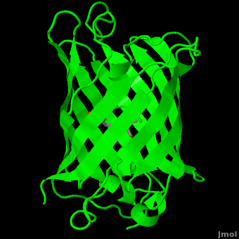
The crystal structure of GFP [1][2] is an eleven-stranded anti-parallel beta-barrel, threaded by an alpha-helix, running up along the axis of the cylinder. In the structure is colored by secondary structure(Alpha Helices and Beta Strands ) to better show the eleven strands and the helices.
.
:
The chromophore is in the distorted alpha-helix that runs along the axis of the can, close to the center of the can-like cylinder.
The chromophore is formed from the tripeptide motif Serine65-Tyrosine66-Glycine67 after translation and folding of GFP. As the GFP protein folds into its native conformation, these three amino acids are forced into a sharp turn, greatly favoring a nucleophilic attack of the amide of Glycine67 on the carbonyl of Serine65, leading to imidazolinone formation by cyclization and dehydration. At this point, GFP is not fluorescent; however, in the presence of molecular oxygen, the α–β bond of Tyrosine66 is subsequently dehydrogenated into conjugation with the imidazolinone, which results in maturation of the GFP chromophore to its fluorescent form [3][4].
The chromophore .
Reference for the Structure
- Ormo M, Cubitt AB, Kallio K, Gross LA, Tsien RY, Remington SJ. Crystal structure of the Aequorea victoria green fluorescent protein. Science. 1996 Sep 6;273(5280):1392-5. PMID:8703075
Related Structures and Topics
- 1ema Aequorea victoria Green Fluorescent Protein
- 1gfl Aequorea victoria Green Fluorescent Protein
- 1b9c Aequorea victoria Green Fluorescent Protein Mutant F99s, M153t And V163a
- GFP featured at the Molecule of the Month series of tutorials by David Goodsell.
Notes and Literature References
- ↑ Ormo M, Cubitt AB, Kallio K, Gross LA, Tsien RY, Remington SJ. Crystal structure of the Aequorea victoria green fluorescent protein. Science. 1996 Sep 6;273(5280):1392-5. PMID:8703075
- ↑ Yang F, Moss LG, Phillips GN Jr. The molecular structure of green fluorescent protein. Nat Biotechnol. 1996 Oct;14(10):1246-51. PMID:9631087 doi:10.1038/nbt1096-1246
- ↑ Heim R, Prasher DC, Tsien RY. Wavelength mutations and posttranslational autoxidation of green fluorescent protein. Proc Natl Acad Sci U S A. 1994 Dec 20;91(26):12501-4. PMID:7809066
- ↑ Cubitt AB, Heim R, Adams SR, Boyd AE, Gross LA, Tsien RY. Understanding, improving and using green fluorescent proteins. Trends Biochem Sci. 1995 Nov;20(11):448-55. PMID:8578587
Additional Literature and Resources
- Tsien RY. The green fluorescent protein. Annu Rev Biochem. 1998;67:509-44. PMID:9759496 doi:10.1146/annurev.biochem.67.1.509
- GFP featured at the Molecule of the Month series of tutorials by David Goodsell.
- The GFP site by Marc Zimmer, Ph. D., at Connecticut College, who authored Glowing Genes: A Revolution In Biotechnology.
Green fluorescent protein (GFP) is a bioluminescent polypeptide consisting of 238 residues isolated from the body of Aequorea victoria jellyfish.[1] GFP converts the blue chemiluminescent of aequorin in the jellyfish into green fluorescent light.[2] In remains unclear why these jellyfish use fluorescence, why green is better than blue, or why they produce a separate protein for green fluorescence as opposed to simply mutating the present aequorin to shift its wavelength,[3] but in the laboratory, GFP can be incorporated into a variety of biological systems in order to function as a marker protein. Since its discovery in 1962, GFP has become a significant contributor to the research of monitoring gene expression, localization, mobility, traffic, interactions between various membrane and cytoplasmic proteins, as well as many others.[4]
History
Aequorea victoria was first discovered and investigated for its bioluminescence by Frank Johnson, who invited Osamu Shimomura to work with him in on a small island not far from British Columbia, where the jellyfish is abundant.[5] Found off the west coast of the United States between British Columbia and central California,[6] the jellyfish was considered a local phenomenon as it would drift in and out of the harbors.[5]
Shimomura was originally looking only to isolate the blue luminescent protein of Aequorea victoria, traditionally thought to be luciferase, but it would soon become apparent that the glow was in fact due to aequorin, a substance related, but slightly varying from luciferase.[4][5] However, the light emitted from aequorin still differed from the light emitted from the wild jellyfish. This quandary led to the discovery of the green fluorescent protein responsible for this disparity, but sufficient amounts of the protein could not be collected for study until 1979. The journey to discover the nature of GFP had begun.[5]
In the 1990’s, Douglas Prasher, Frank Predergast, and co-workers successfully cloned the gene that encoded for GFP. Martin Chalfie further pursued this line of work and was eventually able to express GFP in heterologous systems such as E. coli and C. elegans. Chalfie’s research provided the first evidence that GFP was unique as it did not require the presence of any exogenous substance or cofactor for fluorescence.[4] The lack for the need for a cofactor proved that the cloned GFP gene contained all the information necessary for posttranslational synthesis of the chromophore. [3]
Roger Tsien and co-workers were intrigued by the absence of a necessary cofactor and began to research the structure of GFP and how it relates to its fluorescence. They discovered that a helix within the beta barrel structure of GFP actually contained a fluorophore responsible for fluorescence. In researching its structure, they were able to develop GFP derivatives with improved fluorescence and photo-stability. Shimomura, Chalfie, and Tsien were each recognized for their work involving GFP with the Nobel Prize in 2008.[4] In the time since the work of these three researchers, GFP has been successfully expressed and utilized in bacteria, yeast, slime mold, plants, drosophila fruit flies, zebra-fish, and mammalian cells.[2] Below, mice have had GFP inserted into their genomes for studies in neurology.

Structure
Primary & Secondary Structure
| |||||||||
| 1ema, resolution 1.90Å () | |||||||||
|---|---|---|---|---|---|---|---|---|---|
| Non-Standard Residues: | , | ||||||||
| |||||||||
| |||||||||
| Resources: | FirstGlance, OCA, RCSB, PDBsum | ||||||||
| Coordinates: | save as pdb, mmCIF, xml | ||||||||


Green fluorescent protein () is a 21 kDa protein consisting of 238 residues strung together to form a
of five α-helices and one eleven-stranded β-pleated sheet,[1] where each strand contains nine to thirteen residues each.[7] (To view the primary and secondary structure of GFP, go to [www.ebi.aci.uk].) These β-strands display an almost “seamless symmetry” in which only two of the strands vary in structural content.[8] This β-sheet conforms itself through regular hydrogen bonding into a β-barrel.[2] In GFP, the structure is so regular that of water molecules (red) can be seen following the structure of the barrel.[8] Together with the α-helices at either end of the molecule, a nearly perfect cylinder is produced, 42Å long and 24Å in diameter,[7] creating what is referred to as a “β-can” formation.[8] The short helical segments at either end of the cylinder form “caps” to further protect the interior of the β-barrel.[8] Overall stability is maintained by this β-can structure, helping to resist unfolding from heat and other denaturants.[2]
One can be found running through the central axis of the β-barrel,[4] roughly to the symmetry axis of the barrel.[7] This helix is extremely important as it contains the fluorophore responsible for fluorescence.[2][4] This α-helix in particular is highly stabilized by the many that are made with each strand of the barrel.[9]
The Chromophore
The of GFP is located at the center of the β-barrel with a wild-type excitation peak of 395 nm, and a minor peak at 475 nm (about three times less intense[3]) [2][10][7][8] with extinction coefficients of approximately 30,000 and 7,000 M-1 cm-1, respectively.[2][8] Interestingly, the Aequorea victoria jellyfish utilizes the smaller of the two excitation peaks as pure aequorin emits a light of 470 nm.[3] The relative amplitudes of these two excitation peaks can vary depending on environmental factors and previous illumination.[7] For example, continued excitation leads to a diminution of the 395 nm excitation peak with a reciprocal amplification of the 475 nm peak.[8] Regardless of absorption, the chromophore of GFP emits light of 508 nm.[2][10][7][8]
Three amino residues in the central α-helix constitute the fluorophore of GFP: Ser65Tyr66Gly67 (see left). Tsien et al. discovered that this tri-peptide sequence is post-translationally modified by internal cyclization and oxidation[4] to produce a structure.[2] Studies with E. coli proposed a sequential mechanism for the formation of the fluorophore that was initiated by a rapid cyclization between Ser65 and Gly67 to form an imidazolin-5-one intermediate.[2] This rapid cyclization is carried out via nucleophilic attack of the amino group from Gly67 on the carbonyl group of Ser65 to form a five-membered ring. The loss of water then forms the imidazolin-5-one intermediate.[10] Cyclization is succeeded by a much slower rate-limiting oxygenation of the Tyr66 hydroxybenzyl side chain by atmospheric oxygen (No fluorescence was seen in anaerobically grown E. coli.), resulting in the 4-(p-hydroxybenzylidene)-imidazolidin-5-one stucture.[2][10][8] The double bond that results from this series of reactions results in the linkage of the two π-systems of the rings, forming a larger conjugated system essential for fluorophore stability. [11]

The process is completely auto-catalytic such that there are no known co-factors or enzymatic components required.[2] Despite the stability of the final product, while the chromophore is forming, the environmental temperature cannot drop below 30°C or the yield of viable GFP will decrease substantially.[2][8] This, of course, is not an issue for the protein in nature as the jellyfish is unlikely to encounter waters of this degree in the Pacific Northwest.[3] Such a temperature sensitivity is only relevant during formation as the stability of the final product is maintained through a network of close contacts surrounding the fluorophore.[2] This, however, can and has been used in in pulse-chase experiments in which the GFP-expressing cells are exposed to varying temperatures in place of labeled vs. unlabeled trials.[3]
As the central α-helix is not located directly in the center of the β-barrel, cavities of differing area exist on either side of the chromophore. The larger cavity, consisting of about 135 Å,[7] does not open out to the bulk solvent, but rather houses .[7][12] Had this space not been occupied, it would be expected to considerably destabilize the protein as a whole. The hydrogen bonding created by the presence of the water molecules, however, helps to link the buried of Glu222 and Gln69 that would otherwise be actively polar.[7] Therefore, the water molecules are extremely important in establishing a hydrogen bonding network about the chromophor.[13]
Proteopedia Page Contributors and Editors (what is this?)
Michal Harel, Wayne Decatur, Karsten Theis, Eran Hodis, Laura Carbone, Karl Oberholser, Mark Hoelzer, Joel L. Sussman, Alexander Berchansky, Jaime Prilusky, Joseph M. Steinberger, David Canner
