This old version of Proteopedia is provided for student assignments while the new version is undergoing repairs. Content and edits done in this old version of Proteopedia after March 1, 2026 will eventually be lost when it is retired in about June of 2026.
Apply for new accounts at the new Proteopedia. Your logins will work in both the old and new versions.
Shank protein
From Proteopedia
(Difference between revisions)
| Line 12: | Line 12: | ||
====Shank Oligomerization==== | ====Shank Oligomerization==== | ||
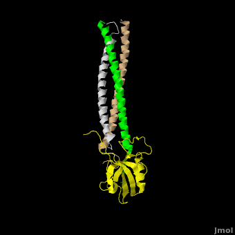
| - | Shank proteins are positioned between scaffolding proteins that are bound to either neurotransmitter receptors or the actin cytoskeleton. This puts Shank proteins in a perfect position to nucleate the underlying structure of the PSD.<ref name="Baron"/> The <scene name='Shank_Family_Proteins/Multimer_opening_single/1'>SAM domain</scene> of Shank-3 can<scene name='Shank_Family_Proteins/Multimer_opening/ | + | Shank proteins are positioned between scaffolding proteins that are bound to either neurotransmitter receptors or the actin cytoskeleton. This puts Shank proteins in a perfect position to nucleate the underlying structure of the PSD.<ref name="Baron"/> The <scene name='Shank_Family_Proteins/Multimer_opening_single/1'>SAM domain</scene> of Shank-3 can<scene name='Shank_Family_Proteins/Multimer_opening/2'>oligomerize</scene> (<scene name='Shank_Family_Proteins/Multimer_opening_alt/2'>Alternate View</scene>) to form large sheets composed of helical fibers stacked side by side. The proposed sheet structure with radially projecting protein interaction domains, is ideal architecture for a protein that must contact both membrane and cytoplasmic components at a synaptic surface.<ref name="Baron"/> Models of this sort validate the importance of Shank-3 as master scaffolding proteins and illustrate how slight mutations can disrupt an entire PSD and synaptic function. |
__NOTOC__ | __NOTOC__ | ||
__NOEDITSECTION__ | __NOEDITSECTION__ | ||
Revision as of 01:20, 29 March 2011
| |||||||||||

Additional Structures of Shank Family Proteins
2fn3 - Crystal Structure of the native Shank SAM domain
2f44 - Crystal Structure of the Zinc-bound Shank SAM domain
1q3o, 1q3p - Crystal structure of the Shank PDZ-ligand complex
References
- ↑ 1.0 1.1 1.2 Park E, Na M, Choi J, Kim S, Lee JR, Yoon J, Park D, Sheng M, Kim E. The Shank family of postsynaptic density proteins interacts with and promotes synaptic accumulation of the beta PIX guanine nucleotide exchange factor for Rac1 and Cdc42. J Biol Chem. 2003 May 23;278(21):19220-9. Epub 2003 Mar 7. PMID:12626503 doi:10.1074/jbc.M301052200
- ↑ 2.0 2.1 2.2 Baron MK, Boeckers TM, Vaida B, Faham S, Gingery M, Sawaya MR, Salyer D, Gundelfinger ED, Bowie JU. An architectural framework that may lie at the core of the postsynaptic density. Science. 2006 Jan 27;311(5760):531-5. PMID:16439662 doi:311/5760/531
- ↑ 3.0 3.1 3.2 Durand CM, Betancur C, Boeckers TM, Bockmann J, Chaste P, Fauchereau F, Nygren G, Rastam M, Gillberg IC, Anckarsater H, Sponheim E, Goubran-Botros H, Delorme R, Chabane N, Mouren-Simeoni MC, de Mas P, Bieth E, Roge B, Heron D, Burglen L, Gillberg C, Leboyer M, Bourgeron T. Mutations in the gene encoding the synaptic scaffolding protein SHANK3 are associated with autism spectrum disorders. Nat Genet. 2007 Jan;39(1):25-7. Epub 2006 Dec 17. PMID:17173049 doi:ng1933
- ↑ 4.0 4.1 4.2 4.3 Bozdagi O, Sakurai T, Papapetrou D, Wang X, Dickstein DL, Takahashi N, Kajiwara Y, Yang M, Katz AM, Scattoni ML, Harris MJ, Saxena R, Silverman JL, Crawley JN, Zhou Q, Hof PR, Buxbaum JD. Haploinsufficiency of the autism-associated Shank3 gene leads to deficits in synaptic function, social interaction, and social communication. Mol Autism. 2010 Dec 17;1(1):15. PMID:21167025 doi:10.1186/2040-2392-1-15
- ↑ Garber K. Neuroscience. Autism's cause may reside in abnormalities at the synapse. Science. 2007 Jul 13;317(5835):190-1. PMID:17626859 doi:10.1126/science.317.5835.190
- ↑ Abu-Elneel K, Liu T, Gazzaniga FS, Nishimura Y, Wall DP, Geschwind DH, Lao K, Kosik KS. Heterogeneous dysregulation of microRNAs across the autism spectrum. Neurogenetics. 2008 Jul;9(3):153-61. Epub 2008 Jun 19. PMID:18563458 doi:10.1007/s10048-008-0133-5
- ↑ 7.0 7.1 7.2 7.3 Im YJ, Kang GB, Lee JH, Park KR, Song HE, Kim E, Song WK, Park D, Eom SH. Structural basis for asymmetric association of the betaPIX coiled coil and shank PDZ. J Mol Biol. 2010 Mar 26;397(2):457-66. Epub 2010 Jan 29. PMID:20117114 doi:10.1016/j.jmb.2010.01.048
Proteopedia Page Contributors and Editors (what is this?)
David Canner, Michal Harel, Alexander Berchansky, Joel L. Sussman
