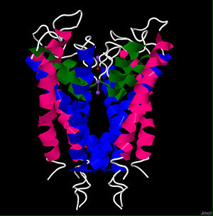
Ion Selectivity
One of the wonders of the potassium channel is its ability to allow the larger K+ ion (1.33Å radius) to pass through the channel, yet simultaneously exclude the smaller Na+ ion (0.95 Å) from entering (Doyle et al., 1998). Interestingly, the channel is 10,000 times more selective for potassium than sodium, yet it shows very little selectivity discrimination between potassium and the next largest alkali metal, rubidium (1.65 Å) (Doyle et al., 1998). This preference for the larger monovalent cations is mediated by a “selectivity filter” that sits at the mouth of the extracellular side of the channel (Doyle et al., 1998). The highly specialized selectivity filter is fully conserved in all known potassium channels, and has a sequence of TVGYG (Morais-Cabral et al., 2001; Doyle et al., 1998). The entire filter is formed when the main-chain carbonyls of the conserved residues from the four subunits point towards the pore to coordinate with the potassium ion (Morais-Cabral et al., 2001). This selectivity filter creates a choke point that provides a very narrow opening to the cavity of the channel (Roux et al., 1999; Doyle et al., 1998). In a seemingly counterintuitive fashion, this choke point actually favors the larger monovalent cations over the smaller ones. The structural determination of the potassium channel showed that this is possible because the carbonyls are perfectly aligned to coordinate with the potassium ion, but the sodium ion is too small to efficiently coordinate with all of the carbonyls from each of the four subunits. The protein-ion coordination allows the selectivity filter to peel away the water molecules that form the tight hydration shell around the potassium ion, effectively reducing the size of the previously solvated cation (Morais-Cabral et al., 2001; Doyle et al., 1998).
The potassium channel has been measured to allow a current of 108 ions/second, meaning the selectivity filter is able to dehydrate a potassium ion in as little as 10 nanoseconds (Morais-Cabral et al., 2001). However, the dehydration of the potassium ion leaves it bound to the carbonyls of the selectivity filter and should, in theory, prevent it from so easily escaping the clutches of the tight coordination to the protein. So what then facilitates such an efficient throughput? From the arrangement of the carbonyls in the resolved structure, it is thought that the selectivity filter is able to bind two ions concomitantly and the electrostatic repulsion between the two adjacent positively charged ions displaces one and pushes it through the channel (Morais-Cabral et al., 2001; Yellen, 2002).
Channel cavity
Before a potassium ion enters the selectivity filter, it passes through an inner pore on the intracellular side of the protein and enters a large, water-filled cavity (Figure 1E) at the bilayer center (Roux et al., 1999; Doyle et al., 1998; Morais-Cabral et al., 2001). This cavity is lined with hydrophobic residues, but holds approximately 50 water molecules (Doyle et al., 1998; Roux et al., 1999). The hydrophobic residues prevent strong binding interaction between the ion and the protein while the water inside the cavity is still able to stabilize the cation as it travels through the channel (Doyle et al., 1998). The C-termini of the four pore helices are angled toward the center of the cavity to assist with the electrostatic stabilization of the ion (Doyle et al., 1998; Yellen, 2002). Each pore helix is a partial dipole, with the C-terminus being slightly negatively charged (Doyle et al., 1998). The slight negative charge points toward the K+ ion while it is in the cavity and confers selectivity of cations over anions (Guidoni et al., 1999)
Voltage gating
The second remarkable aspect of the voltage gated potassium channels is their ability to regulate potassium conductance by sensing and reacting to changes in membrane voltage (Lee et al., 2005). The dependence of potassium channel gating on membrane voltage was first noted in the 1950’s by Hodgkin and Huxley (Hodgkin and Huxley, 1952). They also found that as the channel moved from a low conductance to a high conductance state, a small “gating current” could be measured (Hodgkin and Huxley, 1952). It was initially predicted that this gating current was the result of charged amino acid residues moving through the membrane due to a conformational change in the protein (Cordero-Morales et al., 2006). Under the assumption that the movement of these charges was responsible for the regulation of conductance, mutagenesis experiments were conducted to pinpoint which ionic residues were responsible for gating. Cordero-Morales et al. (2006) found that mutation of Glu71 essentially eliminated the voltage dependence of potassium conductance (Figure 3). A look at the structure of KcsA indicates that this residue is nestled alongside the selectivity filter. The nearby Asp80 residue coordinates with Glu71 to create a carboxyl-carboxylate interaction (Cordero-Morales et al., 2006). It is thought that this carboxyl-carboxylate interaction holds the important residues of the selectivity filter in place. As the voltage changes, this interaction could be disrupted causing a conformational change in the residues of the selectivity filter. Once the residues of the selectivity filter are misaligned, the protein is no longer allows ions to pass through (Cordero-Morales et al., 2006).
Channel Homology
Although the structural determination of the bacterial KcsA channel was no achievement to scoff at, the crown jewel of membrane-protein crystallization was the successful determination of the staggeringly complex Shaker-related mammalian Kv channel (Figure 4) (Miller, 2003a). As opposed to the elegant simplicity of the two-transmembrane helix architecture of the KcsA channel, the Shaker-related potassium channels display a daunting structural complexity that had hindered efforts to crystallize the protein (Miller, 2003b). Earning the Nobel prize in chemistry, Roderick MacKinnon’s lab used a mixture of lipids and detergents to successfully extract and crystallize the Kv channel from a rat in 2003 (Miller, 2003a; Long et al., 2005).
The KcsA channel shares significant sequence and structure homology with the Shaker family of mammalian Kv channels (Doyle et al., 1998). However, each subunit of the Shaker channel consists of 6 helical transmembrane domains (S1-S6) instead of just the two of the KcsA channel (Doyle et al., 1998). The S1-S4 helices are voltage sensing domains and S5-S6 are respectively homologous to the outer and inner helices of the KcsA channel (Yellen, 2002). The selectivity filter structure is strongly conserved between the two proteins, and is thought to work by the same mechanism as well (Long et al., 2007). However, the mechanism of voltage gating in the Shaker related channels is known to occur through a much more complicated mechanism, yet is still not completely understood (Miller, 2003b). For many years it was thought that the S4 helix was located near the core of the protein and oriented parallel to lipids in the membrane. The gating mechanism was thought to involve the S4 helix sliding in and out of the plane of the membrane to somehow cause conformational changes in the rest of the protein (Miller, 2003b; Ahen and Horn, 2004). However, the crystal structure of the channel indicates that the S4 helix is actually oriented perpendicularly to the bilayer, leaving it in a lipid environment (Miller, 2003b; Ahen and Horn, 2004). The S4 helix is known to contain many positively charged residues, meaning it would be energetically unfavorable for the highly polarized helix to exist in the hydrophobic environment that the crystal structure indicates (Miller, 2003; Ahen and Horn, 2004). There remains a highly contentious debate over the merits of the crystal structure and the mechanism of voltage gating in Shaker related potassium channels (Miller, 2003b; Ahen and Horn, 2004).

