Interferon
From Proteopedia
| Line 29: | Line 29: | ||
|<applet load='1HIG.pdb' name='Z' size='300' frame='true' align='right' caption='Interferon Gamma' align='left' scene='Interferon/Ifn_gamma/5'/> | |<applet load='1HIG.pdb' name='Z' size='300' frame='true' align='right' caption='Interferon Gamma' align='left' scene='Interferon/Ifn_gamma/5'/> | ||
|} | |} | ||
| + | <center> | ||
| + | |||
| + | '''Synchronize the three applets showing interferons alpha, beta, and gamma by clicking the checkbox''' | ||
| + | <jmol> | ||
| + | <jmolCheckbox> | ||
| + | <target>A</target> | ||
| + | <!--<scriptWhenChecked>set syncMouse ON;set syncScript OFF;sync jmolAppletB,jmolAppletZ; sync > "set syncMouse | ||
| + | ON;set syncScript OFF"</scriptWhenChecked>--> | ||
| + | <scriptWhenChecked> sync jmolAppletB,jmolAppletZ </scriptWhenChecked> | ||
| + | <scriptWhenUnchecked> sync OFF</scriptWhenUnchecked> | ||
| + | <text> Synchronize</text> | ||
| + | </jmolCheckbox> | ||
| + | </jmol> | ||
| + | </center> | ||
| + | |||
| + | |||
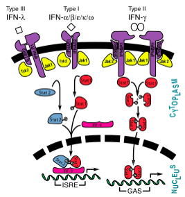
[[Image:InterferonSignalingPathway.png|600px|right|thumb|Interferon JAK-STAT Pathway showing interferons types I, II, and III<ref name="Isaacs">[http://www.jbc.org/content/282/28/20045.full?sid=cbf08059-44d4-4957-8ea7-0351cab9c2ac] Samuel, C.E. "Interferons, Interferon Receptors, Signal Transducer and Transcriptional Activators, and Inteferon Regulatory Factors." ''J Biol Chem'' 2007 282: 20045-20046. First Published on May 14, 2007, doi:10.1074/jbc.R700025200</ref>]] | [[Image:InterferonSignalingPathway.png|600px|right|thumb|Interferon JAK-STAT Pathway showing interferons types I, II, and III<ref name="Isaacs">[http://www.jbc.org/content/282/28/20045.full?sid=cbf08059-44d4-4957-8ea7-0351cab9c2ac] Samuel, C.E. "Interferons, Interferon Receptors, Signal Transducer and Transcriptional Activators, and Inteferon Regulatory Factors." ''J Biol Chem'' 2007 282: 20045-20046. First Published on May 14, 2007, doi:10.1074/jbc.R700025200</ref>]] | ||
Revision as of 18:08, 9 October 2012
Interferons were the first cytokines discovered and were identified by Isaacs and Lindenmann. These proteins were classified as interferons because they interfered with virus growth.[1] The initial experiments performed poorly characterized the interferons, and was based merely on bioactivity. Advances in scientific instrumentation and technique have allowed for greater understanding and visualization of not only the structure but also the mechanisms of the various types of inteferons.[2] The interferons were originally classified as leukocyte (interferon-α), fibroblast (interferon-β), and immmune (interferon-γ), although today they are classified into types I (α, β, ε, κ, ω), II (γ), and III (λ).[1][2]
| |||||||||||
Comparison of three interferons
|
|
|
Synchronize the three applets showing interferons alpha, beta, and gamma by clicking the checkbox

Signaling and Receptor Interactions
The signaling pathways of interferons are interesting as type I interferons share the same receptors IFNAR1 and IFNAR2. Type II interferon-γ has receptors IFNGR1 and IFNGR2, but needs two interferon-γ to signal, as illustrated in the image to the right. Interestingly enough, types I and III act together in the JAK-STAT pathway, while type II acts alone. Interferon-α and -β bind to the same receptors as one another, the affinities with which they bind to IFNAR1 and IFNAR2 differ. While the binding to IFNAR2 is stronger for both in comparison to IFNAR1, interferon-β has a much stronger affinity for IFNAR1 than interferon-α.[9]
Interferon-α to an interferon receptor mainly with helices C and G. There are many within 4 angstroms of one another. These residues could form many , illustrated in white dotted lines. Given that interferon-α does not undergo many structural changes upon binding to interferon receptor II, Quadt-Akabayov et al. have concluded that the binding mechanism is similar to that of a lock and key. Interferons -α and -β interact with a receptor at the cell surface.[10] This receptor has : an , with two disulfide bonds, a , with one disulfide bond, and a . The of the receptor have no secondary structure, allowing for some serious flexibility, leading to .[11]
References
- ↑ 1.0 1.1 1.2 [1] Samuel, C.E. "Interferons, Interferon Receptors, Signal Transducer and Transcriptional Activators, and Inteferon Regulatory Factors." J Biol Chem 2007 282: 20045-20046. First Published on May 14, 2007, doi:10.1074/jbc.R700025200
- ↑ 2.0 2.1 Langer JA, Pestka S. Structure of interferons. Pharmacol Ther. 1985;27(3):371-401. PMID:2413490
- ↑ Quadt-Akabayov SR, Chill JH, Levy R, Kessler N, Anglister J. Determination of the human type I interferon receptor binding site on human interferon-alpha2 by cross saturation and an NMR-based model of the complex. Protein Sci. 2006 Nov;15(11):2656-68. Epub 2006 Sep 25. PMID:17001036 doi:10.1110/ps.062283006
- ↑ Voet, D., Voet, J.G., and C. Pratt. Fundamentals of Biochemistry 3rd Edition. Hoboken, NJ: John Wiley and Sons, 2008. Print.
- ↑ Kudo M. Management of hepatocellular carcinoma: from prevention to molecular targeted therapy. Oncology. 2010 Jul;78 Suppl 1:1-6. Epub 2010 Jul 8. PMID:20616576 doi:10.1159/000315222
- ↑ http://www.uniprot.org/uniprot/P00784
- ↑ Nylander A, Hafler DA. Multiple sclerosis. J Clin Invest. 2012 Apr 2;122(4):1180-8. doi: 10.1172/JCI58649. Epub 2012 Apr 2. PMID:22466660 doi:10.1172/JCI58649
- ↑ Loma I, Heyman R. Multiple sclerosis: pathogenesis and treatment. Curr Neuropharmacol. 2011 Sep;9(3):409-16. PMID:22379455 doi:10.2174/157015911796557911
- ↑ Quadt-Akabayov SR, Chill JH, Levy R, Kessler N, Anglister J. Determination of the human type I interferon receptor binding site on human interferon-alpha2 by cross saturation and an NMR-based model of the complex. Protein Sci. 2006 Nov;15(11):2656-68. Epub 2006 Sep 25. PMID:17001036 doi:10.1110/ps.062283006
- ↑ [2] Samuel, C.E. "Interferons, Interferon Receptors, Signal Transducer and Transcriptional Activators, and Inteferon Regulatory Factors." J Biol Chem 2007 282: 20045-20046. First Published on May 14, 2007, doi:10.1074/jbc.R700025200
- ↑ Chill JH, Quadt SR, Levy R, Schreiber G, Anglister J. The human type I interferon receptor: NMR structure reveals the molecular basis of ligand binding. Structure. 2003 Jul;11(7):791-802. PMID:12842042
3D Structures of interferon
Interferon-α
1itf - hIF 2A – NMR
2hym, 2kz1, 2lag, 3s9d - hIF 2A + IFR α/β
2rh2 – hIF 2B
3se3 - hIF 2B + IFR 1 + IFR 2
3oq3 - hIF 5 + IFR α/β
Interferon-β
1ifa, 1wu3 – IF – mouse
1au1 - hIF
Interferon-γ
1hig – hIF – human
1eku – hIF (mutant)
2rig – IF – rabbit
1rfb, 1d9c – IF – bovine
1fg9, 1fyh, 3bes – hIF + IFR α chain
Interferon-λ
3og4, 3og6 – hIF 1 + IFR
3hhc – hIF 4
Interferon-τ
1b5l – IF
Interferon-ω
3se4 - hIF 1 + IFR 1 + IFR 2
3piv – ZfIF 1 – Zebra fish
3piw - ZfIF 2
Proteopedia Page Contributors and Editors (what is this?)
Michal Harel, Kirsten Eldredge, Alexander Berchansky, Joel L. Sussman, Karl Oberholser, Jaime Prilusky
