User:Erin May/Sandbox 1
From Proteopedia
| Line 34: | Line 34: | ||
| - | === PrP<sup>Sc</sup> | + | === PrP<sup>Sc</sup> === |
<StructureSection load='2rnm' size='300' side='right' caption='Amyloid formation: Human Prion Protein [[2RNM]]' scene=''> | <StructureSection load='2rnm' size='300' side='right' caption='Amyloid formation: Human Prion Protein [[2RNM]]' scene=''> | ||
| - | + | ||
| + | The following residues are shown on the PrP<sup>Sc</sup>: | ||
| + | 129 Val-->Met | ||
| + | R164 to S170 | ||
| + | 200 Glu-->Lys | ||
| + | |||
</StructureSection> | </StructureSection> | ||
Revision as of 06:10, 27 November 2012
Contents |
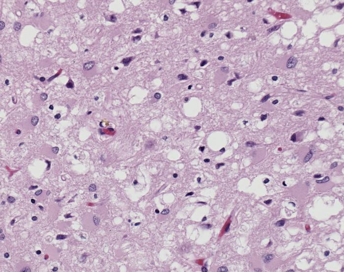
Prions as a disease causing agent

Prions are infectious or genetic misfolded proteins which act as templates upon which properly folded prion protein monomers can aggregate. Prions contain no nucleic acid such as other infectoius molecules or organisms. Human Prion Protein or Major Prion protein, exists as a normal constituent of human cells, found mostly in the brain[2] and is called PrPC.[3] PrPC is composed of mostly helix whereas the infectious form, PrPSc, is composed of high percentage beta sheets.[3]
The diseases prions confer are neurodegenerative disorders which result from the large scale aggregation of these proteins. For more information about the infections related to prions see Transmissible spongiform encephalopathy at Wikipedia.
Unfolding Mechanism
Currently, the mechanism by which a template prion unfolds a the helices of a properly folded prion protein is unknown. Specific residues have been shown to either confer resistance or lend themselves to this unfolding.
PrPC natural monomer
| |||||||||||
PrPSc
| |||||||||||
Dimer Form
| |||||||||||
Reference List
- ↑ Image of Creutzfeldt-Jakob positive brain tissue was obtained from The CDC's Public Health Image Library.
- ↑ Centers for Disease Control and Prevention
- ↑ 3.0 3.1 Prusiner SB. Prions. Proc Natl Acad Sci U S A. 1998 Nov 10;95(23):13363-83. PMID:9811807
- ↑ Lee S, Antony L, Hartmann R, Knaus KJ, Surewicz K, Surewicz WK, Yee VC. Conformational diversity in prion protein variants influences intermolecular beta-sheet formation. EMBO J. 2010 Jan 6;29(1):251-62. Epub 2009 Nov 19. PMID:19927125 doi:10.1038/emboj.2009.333
- ↑ Zhang Y, Swietnicki W, Zagorski MG, Surewicz WK, Sonnichsen FD. Solution structure of the E200K variant of human prion protein. Implications for the mechanism of pathogenesis in familial prion diseases. J Biol Chem. 2000 Oct 27;275(43):33650-4. PMID:10954699 doi:10.1074/jbc.C000483200
