VRC01 gp120 complex
From Proteopedia
| Line 1: | Line 1: | ||
The crystal structure of VRC01 and VRC01-like antibodies are studied to define with characteristics are important in neutralizing HIV-1. (cite!) | The crystal structure of VRC01 and VRC01-like antibodies are studied to define with characteristics are important in neutralizing HIV-1. (cite!) | ||
| - | <Structure load='3NGB' size='500' frame='true' align='left' caption='Trimeric gp120 in complex with VRC01 antibodies' scene='Insert optional scene name here' /> | ||
| - | |||
| - | |||
==Introduction== | ==Introduction== | ||
HIV-1 has a high level of antigenic and genetic diversity. HIV-1 has also evolved mechanisms to evade the humoral immune response. These aspects of HIV-1 have made it difficult to develop a vaccine. After several years of infection, 10 to 25% of HIV-1 infected individuals develop neutralizing antibodies. Some antibodies target the transmembrane gp41 molecules of the HIV-1 viral spike, however most target the surface protein gp120. (Wu) VRC01 and VRC01-like antibodies bind to gp120 and are able to neutralize about 90% of HIV-1 isolates. Structural analysis has shown which characteristics of antibodies are essential to its binding with gp120. (Kwong) Discovery of the structure of these antibodies can help develop an effective HIV-1 vaccine. | HIV-1 has a high level of antigenic and genetic diversity. HIV-1 has also evolved mechanisms to evade the humoral immune response. These aspects of HIV-1 have made it difficult to develop a vaccine. After several years of infection, 10 to 25% of HIV-1 infected individuals develop neutralizing antibodies. Some antibodies target the transmembrane gp41 molecules of the HIV-1 viral spike, however most target the surface protein gp120. (Wu) VRC01 and VRC01-like antibodies bind to gp120 and are able to neutralize about 90% of HIV-1 isolates. Structural analysis has shown which characteristics of antibodies are essential to its binding with gp120. (Kwong) Discovery of the structure of these antibodies can help develop an effective HIV-1 vaccine. | ||
| - | |||
| - | |||
==HIV-1 Neutralization== | ==HIV-1 Neutralization== | ||
HIV-1 enters its host by binding viral gp120, a surface glycoprotein of HIV, to the host cell’s CD4 receptor. This interaction induces conformational changes in gp120. (Wu) This conformational change results in the exposure of a binding site for the co-receptor, usually CCR5 OR CXCR4. (Li) The conformational changes also result in the formation of a pre-hairpin intermediate conformation in which gp41, a transmembrane glycoprotein of HIV, rearranges its molecules so that its N-terminal peptides form a trimer of helices that present a fusion peptide to the target cell. Once fusion occurs between the fusion peptide and the target cell membrane, HIV is able to enter and infect the target cell. (Tran) VRC01 binds to CD4’s binding site on gp120, preventing the CD4 receptor from binding to HIV and infecting the cell. (Wu). | HIV-1 enters its host by binding viral gp120, a surface glycoprotein of HIV, to the host cell’s CD4 receptor. This interaction induces conformational changes in gp120. (Wu) This conformational change results in the exposure of a binding site for the co-receptor, usually CCR5 OR CXCR4. (Li) The conformational changes also result in the formation of a pre-hairpin intermediate conformation in which gp41, a transmembrane glycoprotein of HIV, rearranges its molecules so that its N-terminal peptides form a trimer of helices that present a fusion peptide to the target cell. Once fusion occurs between the fusion peptide and the target cell membrane, HIV is able to enter and infect the target cell. (Tran) VRC01 binds to CD4’s binding site on gp120, preventing the CD4 receptor from binding to HIV and infecting the cell. (Wu). | ||
| - | <StructureSection load='3SE9' color='structure' size='500' frame='true' align='left' caption=' | + | <StructureSection load='3SE9' color='structure' size='500' frame='true' align='left' caption='VRC01 in complex with gp120' > |
| Line 18: | Line 13: | ||
==Structural Features== | ==Structural Features== | ||
| - | <u> | + | <u>Similarities to CD4 in complex with gp120</u>. ToxT belongs to a family of transcriptional regulators headed by and known as AraC.<ref name="structure">PMID: 20133655</ref> The AraC family is characterized by a 100 amino acid region of sequence similarity that forms a <scene name='ToxT_Transcriptional_Regulator_in_Vibrio_cholerae/Two_hth_domains/1'>DNA-binding domain</scene> with two helix-turn-helix motifs (one on either side of the black linker). <ref name="arac">PMID: 11282467</ref> This DNA binding domain is composed of seven alpha helices. HTH1 is composed of alpha helices five and six, while HTH2 is composed of alpha helices eight and nine. The two HTH regions are linked by the very polar alpha helix seven(shown in black). The overall domain is located at the C-terminus.<ref name="structure">PMID: 20133655</ref> Assuming ToxT is similar in mechanism to other AraC proteins, helix six from HTH1 and helix nine from HTH2 become aligned with the help of helix seven. Helix seven is positioned to attach to the N terminal binding pocket(the polar linking region) to allow binding to major consecutive grooves of target DNA (specific promoters for virulence genes).<ref name="structure">PMID: 20133655</ref>[http://www.pnas.org/content/107/7/2860/F3.large.jpg]. The conformation of helix seven is dependent on the ligand bound. |
<br/> | <br/> | ||
<br/> | <br/> | ||
| - | <u> | + | <u>Similarities to other antibodies in complex with gp120</u>. <scene name='ToxT_Transcriptional_Regulator_in_Vibrio_cholerae/Barrel/1'>A nine-stranded beta sheet sandwich</scene> or "jelly-roll" with three other alpha helices (overall making up the <scene name='ToxT_Transcriptional_Regulator_in_Vibrio_cholerae/N-terminus/1'>N-terminus</scene>) contain a <scene name='ToxT_Transcriptional_Regulator_in_Vibrio_cholerae/Binding_pocket/1'>binding pocket</scene>. This is made from several residues from the N-terminus (Y12, Y20, F22, L25, I27, K31, F33, L61, F69, L71, V81, and V83), and a few from the C-terminus (I226, K230, M259, V261, Y266, and M269). This pocket contains a sixteen-carbon fatty acid positioned in a conformation such that its negatively charged carboxylate group forms salt bridges between K31 of the N-terminal domain, and K230 from the C-terminal domain. The pocket is highly hydrophobic, and has a known volume of 780.9 Angstroms.<ref name="structure">PMID: 20133655</ref> This pocket contains a ligand: <scene name='ToxT_Transcriptional_Regulator_in_Vibrio_cholerae/Binding_pocket/2'>cis-palmitoleate</scene> <ref name="structure">PMID: 20133655</ref> which appears to have a negative effect on virulence when present in vitro. The <i>cis</i>-palmitoleate forms <scene name='ToxT_Transcriptional_Regulator_in_Vibrio_cholerae/Salt_bridges_pam/1'>salt bridges</scene> with residues K31 and K230 (for detail, see Figure 1B of: [http://www.pnas.org/content/107/7/2860/F1.large.jpg]). This unsaturated fatty acid, like other UFAs,[http://en.wikipedia.org/wiki/Fatty_acid#Unsaturated_fatty_acids] tend to inhibit genes under the control of ToxT. |
Specifically, the <i>cis</i>-palmitoleate (PAM) appears to change ToxT's conformation, and thus lower its ability to bind DNA and form dimers.<ref name="structure">PMID: 20133655</ref> The presence of UFAs is associated with being in the lumen of the intestine during the bacterial infection. PAM brings K31 and K230 together from either end of the protein, and essentially closes off ToxT. K230 is at the end of helix seven, and binding to K31 causes helix six to be pulled into an unfavorable conformation that deters DNA binding. In lower concentration of fatty acids, ie: after penetrating the intestine's mucus, PAM is in lower concentration. At this point, charge-charge repulsion between K31 and K230 leads to a destabilization of the closed conformation of ToxT. This repulsion prompts the opening of the N and C terminal domains. The freedom of helices six and seven to find a favorable configuration allows DNA binding to occur.<ref name="structure">PMID: 20133655</ref> | Specifically, the <i>cis</i>-palmitoleate (PAM) appears to change ToxT's conformation, and thus lower its ability to bind DNA and form dimers.<ref name="structure">PMID: 20133655</ref> The presence of UFAs is associated with being in the lumen of the intestine during the bacterial infection. PAM brings K31 and K230 together from either end of the protein, and essentially closes off ToxT. K230 is at the end of helix seven, and binding to K31 causes helix six to be pulled into an unfavorable conformation that deters DNA binding. In lower concentration of fatty acids, ie: after penetrating the intestine's mucus, PAM is in lower concentration. At this point, charge-charge repulsion between K31 and K230 leads to a destabilization of the closed conformation of ToxT. This repulsion prompts the opening of the N and C terminal domains. The freedom of helices six and seven to find a favorable configuration allows DNA binding to occur.<ref name="structure">PMID: 20133655</ref> | ||
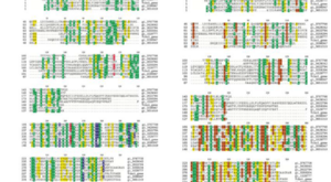
<br/> [[Image:MSA.png|center|300px|thumb| MSA [[1xtc]]]] | <br/> [[Image:MSA.png|center|300px|thumb| MSA [[1xtc]]]] | ||
<br/> | <br/> | ||
| - | <u> | + | <u>Other features</u>. Though the structure shown is a monomer with two overall domains (N-terminal and C-terminal), ToxT tends to form a dimer.<ref name="dimerization">PMID: 21415495 |
</ref> The preferred state of ToxT varies between promoters, but binding to the <i>ctx</i> promoter to generate cholera toxin appears to be possible only in the dimer form.<ref name="virstatin">PMID:17283330</ref>ToxT binds to thirteen base pair sequences (can be single, direct, or inverted repeats) called toxboxes in order to activate their respective promoters.[http://www.sigwiki.info/wiki/Signature:ToxBox] | </ref> The preferred state of ToxT varies between promoters, but binding to the <i>ctx</i> promoter to generate cholera toxin appears to be possible only in the dimer form.<ref name="virstatin">PMID:17283330</ref>ToxT binds to thirteen base pair sequences (can be single, direct, or inverted repeats) called toxboxes in order to activate their respective promoters.[http://www.sigwiki.info/wiki/Signature:ToxBox] | ||
Revision as of 07:13, 27 November 2012
The crystal structure of VRC01 and VRC01-like antibodies are studied to define with characteristics are important in neutralizing HIV-1. (cite!)
Contents |
Introduction
HIV-1 has a high level of antigenic and genetic diversity. HIV-1 has also evolved mechanisms to evade the humoral immune response. These aspects of HIV-1 have made it difficult to develop a vaccine. After several years of infection, 10 to 25% of HIV-1 infected individuals develop neutralizing antibodies. Some antibodies target the transmembrane gp41 molecules of the HIV-1 viral spike, however most target the surface protein gp120. (Wu) VRC01 and VRC01-like antibodies bind to gp120 and are able to neutralize about 90% of HIV-1 isolates. Structural analysis has shown which characteristics of antibodies are essential to its binding with gp120. (Kwong) Discovery of the structure of these antibodies can help develop an effective HIV-1 vaccine.
HIV-1 Neutralization
HIV-1 enters its host by binding viral gp120, a surface glycoprotein of HIV, to the host cell’s CD4 receptor. This interaction induces conformational changes in gp120. (Wu) This conformational change results in the exposure of a binding site for the co-receptor, usually CCR5 OR CXCR4. (Li) The conformational changes also result in the formation of a pre-hairpin intermediate conformation in which gp41, a transmembrane glycoprotein of HIV, rearranges its molecules so that its N-terminal peptides form a trimer of helices that present a fusion peptide to the target cell. Once fusion occurs between the fusion peptide and the target cell membrane, HIV is able to enter and infect the target cell. (Tran) VRC01 binds to CD4’s binding site on gp120, preventing the CD4 receptor from binding to HIV and infecting the cell. (Wu).
| |||||||||||
HIV Prevention Research
In 2003, Veazey and fellow researchers found that early broadly neutralizing antibodies had microbicide potential by using a monkey cell as the model. The microbicide used on these monkeys consisted of b12, a broadly neutralizing antibody. These monkeys were challenged with SHIV, simian-human immunodeficiency virus, through the vagina. Only three of the twelve monkeys became infected. It was also found that the protection against HIV lasts for up to two hours. (Veazy)These results show that microbicides containing antibodies are effective at preventing HIV in monkeys.
A similar experiment was done in 2012; it used humanized mouse models called RAG-hu mice, which contained human target cells. Results show that seven out of nine mice that were administered the VRC01 antibody and all mice that were given a cocktail containing four broadly neutralizing antibodies as a topical gel were protected against HIV-1. These results showed that broadly neutralizing antibodies could be used as a topical microbicide to prevent vaginal transmission of HIV and that a combination of antibodies can provide better protection against HIV. When the VRC01 antibody and the broadly neutralizing antibody cocktail were administered to the humanized mice via the intravenous route, none of the mice were infected with SHIV. (Veselinovic)
References
Proteopedia Page Contributors and Editors (what is this?)
Amanda Valdiosera, Michal Harel, Chris Casey, Alexander Berchansky

