This old version of Proteopedia is provided for student assignments while the new version is undergoing repairs. Content and edits done in this old version of Proteopedia after March 1, 2026 will eventually be lost when it is retired in about June of 2026.
Apply for new accounts at the new Proteopedia. Your logins will work in both the old and new versions.
Vitamin D receptor
From Proteopedia
| Line 2: | Line 2: | ||
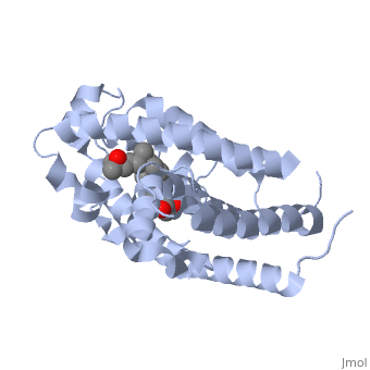
<StructureSection load='1db1' size='350' side='right' caption='Structure of human vitamin D receptor ligand-binding domain complex with vitamin D (PDB entry [[1db1]])' scene=''> | <StructureSection load='1db1' size='350' side='right' caption='Structure of human vitamin D receptor ligand-binding domain complex with vitamin D (PDB entry [[1db1]])' scene=''> | ||
| - | '''Vitamin D receptor''' (VDR) is a transcription factor. Upon binding to vitamin D, VDR forms a heterodimer with retinoid-X receptor and binds to hormone response receptors on DNA causing gene expression. | + | '''Vitamin D receptor''' (<scene name='56/562378/Vit_d_receptor_3m7r/3'>VDR</scene>) is a transcription factor. Upon binding to vitamin D, VDR forms a heterodimer with retinoid-X receptor and binds to hormone response receptors on DNA causing gene expression. The <scene name='56/562378/Vit_d_receptor_ligand/1'>vitamin D hormone</scene> (in green) binds to receptors in its target cells, controlling the synthesis of many different proteins involved in calcium transport and utilization. VDR contains two domains: a <scene name='56/562378/Lbd/1'>ligand binding domain (LBD)</scene> that binds to the hormone (grey) and <scene name='56/562378/Dbd/2'>DNA-binding domain (DBD)</scene> that binds to DNA. (Green and blue are two same VDR structures). It pairs up with a similar protein, 9-cis retinoic acid receptor (RXR), and together they bind to the DNA, activating synthesis in some cases and repressing it in others. |
| + | |||
| + | |||
| + | ===Crystal Structure of the nuclear receptor for Vitamnin D complexed to Vitamin D=== | ||
| + | |||
| + | {{ABSTRACT_PUBMED_10678179}} | ||
| + | |||
| + | |||
| + | ==Disease== | ||
| + | [[http://www.uniprot.org/uniprot/VDR_HUMAN VDR_HUMAN]] Defects in VDR are the cause of rickets vitamin D-dependent type 2A (VDDR2A) [MIM:[http://omim.org/entry/277440 277440]]. A disorder of vitamin D metabolism resulting in severe rickets, hypocalcemia and secondary hyperparathyroidism. Most patients have total alopecia in addition to rickets.<ref>PMID:2849209</ref><ref>PMID:8381803</ref><ref>PMID:1652893</ref><ref>PMID:2177843</ref><ref>PMID:8106618</ref><ref>PMID:8392085</ref><ref>PMID:7828346</ref><ref>PMID:8675579</ref><ref>PMID:8961271</ref><ref>PMID:9005998</ref> | ||
| + | |||
| + | |||
| + | ==Function== | ||
| + | Vitamin D plays an essential role in regulating the levels of calcium and phosphate in the body. It is converted into a hormone that is secreted by the kidneys and travels through the body. It has major effects on intestinal cells, where it helps control the uptake of calcium, and bone cells, where it helps control the formation and maintenance of the skeleton. | ||
| + | |||
| + | [[http://www.uniprot.org/uniprot/VDR_HUMAN VDR_HUMAN]] Nuclear hormone receptor: Transcription factor that mediates the action of vitamin D3 by controlling the expression of hormone sensitive genes. Regulates transcription of hormone sensitive genes via its association with the WINAC complex, a chromatin-remodeling complex. Recruited to promoters via its interaction with the WINAC complex subunit BAZ1B/WSTF, which mediates the interaction with acetylated histones, an essential step for VDR-promoter association. Plays a central role in calcium homeostasis.<ref>PMID:16252006</ref><ref>PMID:10678179</ref><ref>PMID:15728261</ref><ref>PMID:16913708</ref> | ||
| + | |||
| + | |||
| + | ==Mutation== | ||
| + | <StructureSection load='VDRmutation1.pdb' size='350' side='right' caption='Mutation of Vitamin D Receptor' scene=''>In the article, "Phosphorylation of the Human Vitamin D receptor by Protein Kinase C" by Hsieh, J. et al, they presented their research on the mutation of serine to glycine and aspartic acid. They mentioned that amino acids like serine and threonine kinase plays a crucial role in signal transduction pathways drawn out by variety of growth factors, hormones, and neurotransmitters. When <scene name='56/562378/Serine_final/1'>serine</scene> is mutated it is replaced with a <scene name='56/562378/Glycine_final/1'>glycine</scene> which results in an inhibition of transcriptional activation. When transcription is inhibited it results in p53 accumulation, which activates and promotes p53 translocation into mitochondria leading to apoptosis. Transcription inhibition is useful in cancer patients and so can be used as treatment option. These are the outcomes of the mutation, with the research still in the process to find the potential cure for tumors. | ||
| + | |||
| + | |||
| + | |||
| + | <scene name='56/562378/Serine_final/1'>Serine</scene> is replaced with <scene name='56/562378/Asparticacid_final/1'>aspartic acid</scene> when mutated creating a negative charge. The negative charge at the residue inhibits DNA binding which cause a down – regulation of VDR activity. VDR needs DNA binding in order for it to be activated which is only possible with a serine residue. Research is still continuing to find a therapeutic cause for this mutation. | ||
| + | </StructureSection> | ||
| + | |||
| + | |||
| + | ==Crystal structure of the human VDR ligand binding domain bound to the synthetic agonist compound 2alpha-methyl-AMCR277A(C23S)== | ||
| + | <StructureSection load='3a3z' size='350' side='right' caption='Synthetic agonist (PDB entry [[3a3z]])' scene=''>The vitamin D nuclear receptor is a ligand-dependent transcription factor that controls multiple biological responses such as cell proliferation, immune responses, and bone mineralization. Numerous 1 alpha,25(OH)(2)D(3) analogues, which exhibit low calcemic side effects and/or antitumoral properties, have been synthesized. In the article, "Structure-function relationships and crystal structures of the vitamin D receptor bound 2 alpha-methyl-(20S,23S)- and 2 alpha-methyl-(20S,23R)-epoxymethano-1 alpha,25-dihydroxyvitamin D3" by Antony, P. et al, they showed that <scene name='56/562378/3a3z/1'>the synthetic analogue (20S,23S)-epoxymethano-1alpha,25-dihydroxyvitamin D(3) (2a)</scene> acts as a 1alpha,25(OH)(2)D(3) superagonist and exhibits both antiproliferative and prodifferentiating properties in vitro. Using this information and on the basis of the crystal structures of human VDR ligand binding domain (hVDR LBD) bound to 1alpha,25(OH)(2)D(3), 2alpha-methyl-1alpha,25(OH)(2)D(3), or 2a, we designed a novel analogue, 2alpha-methyl-(20S,23S)-epoxymethano-1alpha,25-dihydroxyvitamin D(3) (4a), in order to increase its transactivation potency. Here, we solved the crystal structures of the hVDR LBD in complex with the 4a (C23S) and its epimer 4b (C23R) and determined their correlation with specific biological outcomes. | ||
| + | </StructureSection> | ||
| + | |||
| + | |||
| + | ==About this Structure== | ||
| + | [[1db1]] is a 1 chain structure with sequence from [http://en.wikipedia.org/wiki/Homo_sapiens Homo sapiens]. Full crystallographic information is available from [http://oca.weizmann.ac.il/oca-bin/ocashort?id=1DB1 OCA]. On right hand side is Structure of human vitamin D receptor ligand-binding domain complex with vitamin D (PDB entry [[1db1]]). | ||
| + | |||
==3D structures of vitamin D receptor== | ==3D structures of vitamin D receptor== | ||
Revision as of 11:59, 3 February 2014
| |||||||||||
Contents |
Crystal structure of the human VDR ligand binding domain bound to the synthetic agonist compound 2alpha-methyl-AMCR277A(C23S)
| |||||||||||
About this Structure
1db1 is a 1 chain structure with sequence from Homo sapiens. Full crystallographic information is available from OCA. On right hand side is Structure of human vitamin D receptor ligand-binding domain complex with vitamin D (PDB entry 1db1).
3D structures of vitamin D receptor
Updated on 03-February-2014
Vitamin D receptor ligand-binding domain
3m7r - hVDR LBD (mutant) – human
1db1 – hVDR LBD + vitamin D
1s0z, 1s19, 3a78, 4g2i - hVDR LBD + vitamin D derivative
3ogt, 2ham, 2har, 2has, 2hb7, 2hb8, 3p8x, 3az1, 3az2, 3az3, 3tkc - hVDR LBD + vitamin D analog
3auq, 3aur, 3kpz - hVDR LBD (mutant) + vitamin D analog
3a2i, 3a2j, 3b0t, 3ax8, 3vhw - hVDR LBD (mutant) + vitamin D derivative
3a3z, 3a40 - hVDR LBD + agonist
1ie8, 1ie9, 3cs4, 3cs6, 1txi – hVDR LBD + superagonist
3w5q, 3w5r, 3w5t - hVDR LBD + lithocholic acid derivative
Vitamin D receptor LBD complex with peptide
1rjk, 1rk3, 1rkg, 1rkh, 2o4j, 2o4r – rVDR LBD (mutant) + peroxisome proliferator-activated receptor peptide – rat
2zl9, 2zla, 2zlc - rVDR LBD + coactivator peptide DRIP + vitamin D analog
3vrt, 3vru, 3vrv, 3vrw - rVDR LBD (mutant) + coactivator peptide DRIP + vitamin D analog
2zfx, 3a2h, 2zxm, 2zxn - rVDR LBD + coactivator peptide DRIP
3aun, 2zmh, 2zmi, 2zmj, 3afr, 3vjs, 3vjt - rVDR LBD (mutsant) + coactivator peptide DRIP
2hbh - zVDR LBD + steroid receptor coactivator 1 peptide – zebrafish
2hc4 - zVDR LBD + steroid receptor coactivator 1 peptide + vitamin D
2hcd - zVDR LBD + steroid receptor coactivator 1 peptide
Vitamin D receptor DNA-binding domain
1kb2 – hVDR DBD + osteopontin response element DNA
1kb4 – hVDR DBD + DR3 response element DNA
1ynw – hVDR DBD (mutant) + DR3 response element DNA
1kb6 – hVDR DBD + osteocalcin response element DNA
</StructureSection>
Proteopedia Page Contributors and Editors (what is this?)
Michal Harel, Alexander Berchansky, Jaime Prilusky, Isita Amin
