We apologize for Proteopedia being slow to respond. For the past two years, a new implementation of Proteopedia has been being built. Soon, it will replace this 18-year old system. All existing content will be moved to the new system at a date that will be announced here.
Introduction to Evolutionary Conservation
From Proteopedia
(Difference between revisions)
(→Locations of Mutations in 3D Model) |
|||
| Line 1: | Line 1: | ||
| + | <StructureSection load='' size='350' side='right' scene='Introduction_to_Evolutionary_Conservation/Conservation/1' caption='MeCp2 protein bound to DNA (crystal structure [[3c2i]]). Conservation calculated by [[ConSurfDB_vs._ConSurf|ConSurf-DB]].'> | ||
| + | |||
Mutations occur spontaneously in each generation, randomly changing an amino acid here and there in a protein. Individuals with mutations that impair critical functions of proteins may have resulting problems that make them less able to reproduce. Harmful mutations are lost from the gene pool because the individuals carrying them reproduce less effectively. Since the harmful mutations are lost, the amino acids critical for the function of a protein are '''conserved'''. In contrast, harmless (or very rare beneficial) mutations are kept in the gene pool, producing '''variability''' in non-critical amino acids. | Mutations occur spontaneously in each generation, randomly changing an amino acid here and there in a protein. Individuals with mutations that impair critical functions of proteins may have resulting problems that make them less able to reproduce. Harmful mutations are lost from the gene pool because the individuals carrying them reproduce less effectively. Since the harmful mutations are lost, the amino acids critical for the function of a protein are '''conserved'''. In contrast, harmless (or very rare beneficial) mutations are kept in the gene pool, producing '''variability''' in non-critical amino acids. | ||
| Line 74: | Line 76: | ||
===Locations of Mutations in 3D Model=== | ===Locations of Mutations in 3D Model=== | ||
| - | <Structure load='' size='350' frame='true' align='right' caption='MeCp2 protein bound to DNA (crystal structure [[3c2i]]). Conservation calculated by [[ConSurfDB_vs._ConSurf|ConSurf-DB]].' scene='Introduction_to_Evolutionary_Conservation/Conservation/1' /> | ||
The positions of <font color='#c000a8'>'''conserved Arg133'''</font> and <font color='00a0a0'>'''variable Glu143'''</font> are highlighted with <span style="background:black; color:yellow;">''' yellow '''</span> halos (<scene name='Introduction_to_Evolutionary_Conservation/Conservation/1'>restore initial scene</scene>). You can see that <font color='#c000a8'>'''conserved Arg133'''</font> is in intimate contact with the <font color='#d46a42'>'''DNA'''</font>, while <font color='00a0a0'>'''variable Glu143'''</font> is on the surface, and remote from the contact with the <font color='#d46a42'>'''DNA'''</font>. | The positions of <font color='#c000a8'>'''conserved Arg133'''</font> and <font color='00a0a0'>'''variable Glu143'''</font> are highlighted with <span style="background:black; color:yellow;">''' yellow '''</span> halos (<scene name='Introduction_to_Evolutionary_Conservation/Conservation/1'>restore initial scene</scene>). You can see that <font color='#c000a8'>'''conserved Arg133'''</font> is in intimate contact with the <font color='#d46a42'>'''DNA'''</font>, while <font color='00a0a0'>'''variable Glu143'''</font> is on the surface, and remote from the contact with the <font color='#d46a42'>'''DNA'''</font>. | ||
| Line 86: | Line 87: | ||
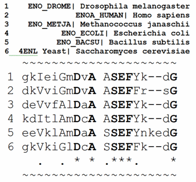
Evolutionary conservation in proteins is identified by aligning the amino acid sequences of proteins with the same function from different taxa (orthologs). As an example, we'll use the glycolytic enzyme '''enolase''', present in a wide range of taxa. Take a quick look to get an impression of a [[Enolase multiple sequence alignment|multiple sequence alignment for ~400 amino acids in enolase]] for taxa ranging from eubacteria and archaebacteria through yeast, insects, and humans. In the full [[Enolase multiple sequence alignment|multiple sequence alignment]] is one <span style="background:pink;"> segment highlighted in pink </span>. This segment is enlarged below. | Evolutionary conservation in proteins is identified by aligning the amino acid sequences of proteins with the same function from different taxa (orthologs). As an example, we'll use the glycolytic enzyme '''enolase''', present in a wide range of taxa. Take a quick look to get an impression of a [[Enolase multiple sequence alignment|multiple sequence alignment for ~400 amino acids in enolase]] for taxa ranging from eubacteria and archaebacteria through yeast, insects, and humans. In the full [[Enolase multiple sequence alignment|multiple sequence alignment]] is one <span style="background:pink;"> segment highlighted in pink </span>. This segment is enlarged below. | ||
| - | < | + | <left> |
[[Image:Msa3d 4enl bw.gif|400 px]]<br> | [[Image:Msa3d 4enl bw.gif|400 px]]<br> | ||
[[Image:Msa3d bw key.gif|400 px]] | [[Image:Msa3d bw key.gif|400 px]] | ||
| - | </ | + | </left> |
By comparing the amino acids in each column, you will find that some positions are 100% identical (conserved) between taxa. These amino acids are in '''BOLD UPPER CASE''' and indicated by an asterisk (*) at the bottom of the column. | By comparing the amino acids in each column, you will find that some positions are 100% identical (conserved) between taxa. These amino acids are in '''BOLD UPPER CASE''' and indicated by an asterisk (*) at the bottom of the column. | ||
| Line 103: | Line 104: | ||
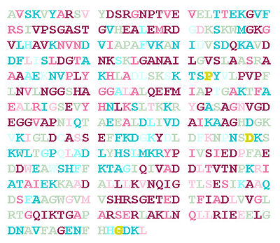
When ConSurf's colors are applied to the 436 amino acids in the sequence of enolase (based on a multiple sequence alignment containing 150 sequences), this is the result: | When ConSurf's colors are applied to the 436 amino acids in the sequence of enolase (based on a multiple sequence alignment containing 150 sequences), this is the result: | ||
[[Image:4enl consurf150 sequence wb.jpg|400 px|left]] | [[Image:4enl consurf150 sequence wb.jpg|400 px|left]] | ||
| - | + | {{Clear}} | |
Notice that the conserved residues are scattered around the sequence with no obvious pattern. However, when the same colors are applied to the amino acids in the 3D structure (<scene name='Introduction_to_Evolutionary_Conservation/Enolase_with_consurf_colors/1'>restore initial scene</scene>), they form a conserved patch around the catalytic site (marked with a <span style="background:black; color:#00ff00;">''' zinc ion colored green '''</span>. | Notice that the conserved residues are scattered around the sequence with no obvious pattern. However, when the same colors are applied to the amino acids in the 3D structure (<scene name='Introduction_to_Evolutionary_Conservation/Enolase_with_consurf_colors/1'>restore initial scene</scene>), they form a conserved patch around the catalytic site (marked with a <span style="background:black; color:#00ff00;">''' zinc ion colored green '''</span>. | ||
| Line 111: | Line 112: | ||
{{Clear}} | {{Clear}} | ||
| - | + | </StructureSection> | |
| + | __NOTOC__ | ||
==See Also== | ==See Also== | ||
*[[How to see conserved regions]] | *[[How to see conserved regions]] | ||
Revision as of 07:35, 12 May 2014
| |||||||||||
See Also
Notes and References
- ↑ MECP2 article in the National Library of Medicine's Genetic Home Reference
- ↑ Advantageous variability will be seen in these cases: 5hmg, 2vaa, 3hi6.


