We apologize for Proteopedia being slow to respond. For the past two years, a new implementation of Proteopedia has been being built. Soon, it will replace this 18-year old system. All existing content will be moved to the new system at a date that will be announced here.
Sandbox Reserved 967
From Proteopedia
(Difference between revisions)
| Line 9: | Line 9: | ||
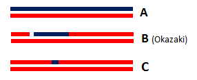
There are two types of RNase H (RNases H1 and RNases H2) classified according to their sequence conservation and substrate preference. Currently, three types of RNA/DNA hybrids are known: simple RNA/DNA duplexes ('''Figure 1A'''), RNA•DNA/DNA hybrids ('''Figure 1B'''), and DNA•RNAfew•DNA/DNA hybrids ('''Figure 1C'''). RNases H2 is totally able to cleave a single ribonucleotide embedded in a double strand DNA (DNA• RNAfew •DNA/DNA type) when RNases H1 require at least 4 ribonucleotides. This ability and their high expression in proliferating cells suggest that RNases H2 are involved in DNA repair and replication. | There are two types of RNase H (RNases H1 and RNases H2) classified according to their sequence conservation and substrate preference. Currently, three types of RNA/DNA hybrids are known: simple RNA/DNA duplexes ('''Figure 1A'''), RNA•DNA/DNA hybrids ('''Figure 1B'''), and DNA•RNAfew•DNA/DNA hybrids ('''Figure 1C'''). RNases H2 is totally able to cleave a single ribonucleotide embedded in a double strand DNA (DNA• RNAfew •DNA/DNA type) when RNases H1 require at least 4 ribonucleotides. This ability and their high expression in proliferating cells suggest that RNases H2 are involved in DNA repair and replication. | ||
| - | [[Image: | + | [[Image:ThreetypesofRNA-DNAhybrids.png|300px|left|thumb| '''Figure 1''' : Three types of RNA/DNA hybrids]] |
| - | + | ||
| - | '' | + | |
Indeed, ribonucleotides are wrongly incorporated into DNA during DNA replication at a frequency of about 2 ribonucleotides per kb. With such frequency, these errors are by far the most abundant threat of DNA damaging. Hence, a correction is essential to the preservation of DNA integrity: the most common correction mechanism involves RNases H2 and is called Ribonucleotide Excision Repair (RER). The incorporation of ribonucleotides in DNA produce DNA•RNAfew•DNA/DNA hybrids from which the few misincorporated ribonucleotides can be removed by an RNase H2. | Indeed, ribonucleotides are wrongly incorporated into DNA during DNA replication at a frequency of about 2 ribonucleotides per kb. With such frequency, these errors are by far the most abundant threat of DNA damaging. Hence, a correction is essential to the preservation of DNA integrity: the most common correction mechanism involves RNases H2 and is called Ribonucleotide Excision Repair (RER). The incorporation of ribonucleotides in DNA produce DNA•RNAfew•DNA/DNA hybrids from which the few misincorporated ribonucleotides can be removed by an RNase H2. | ||
| Line 25: | Line 23: | ||
=== A heteromeric complex === | === A heteromeric complex === | ||
| - | It has been shown that the Mammalian RNase complex is a heteromeric complex formed by 3 distinct proteins: | + | It has been shown that the Mammalian RNase complex is a heteromeric complex formed by 3 distinct proteins: H2A, H2B and H2C. H2A protein is the catalytic subunit and H2B/H2C proteins are auxiliary subunits: they are structural domains that facilitate cohesion of the complex. |
The first domain structure of the complex, H2A, contains 301 amino acids, almost as H2B protein which computes 308 amino acids. H2C protein is the smallest subunit: it only has 166 amino acids. | The first domain structure of the complex, H2A, contains 301 amino acids, almost as H2B protein which computes 308 amino acids. H2C protein is the smallest subunit: it only has 166 amino acids. | ||
Each of these proteins adopts various secondary structures with β-strands and α-helices: | Each of these proteins adopts various secondary structures with β-strands and α-helices: | ||
| Line 52: | Line 50: | ||
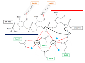
The active site of the catalytic H2A protein is located in a cleft near one end of the complex and formed by strands β2 and 5, and helices α4 and α5. The active site contains four catalytic amino acids strictly conserved in RNases H2: <scene name='60/604486/Site_actif/1'>Asp34, Glu35, Asp142 and Asp170</scene>. These amino acids are essential for the coordination of the 2 divalent metal ions implicated in the stabilisation of reaction intermediates and transition states. In vitro, these ions can be Mg++, Mn++ or Zn++ but the native enzyme is likely to contain Mg++. | The active site of the catalytic H2A protein is located in a cleft near one end of the complex and formed by strands β2 and 5, and helices α4 and α5. The active site contains four catalytic amino acids strictly conserved in RNases H2: <scene name='60/604486/Site_actif/1'>Asp34, Glu35, Asp142 and Asp170</scene>. These amino acids are essential for the coordination of the 2 divalent metal ions implicated in the stabilisation of reaction intermediates and transition states. In vitro, these ions can be Mg++, Mn++ or Zn++ but the native enzyme is likely to contain Mg++. | ||
| - | ''' | + | [[Image:ProposedmechanismforOkazaki.png|300px|left|thumb| '''Figure 2''' : Proposed mechanism for Okazaki fragment processing (not a concerted mechanism)]] |
The hydrolysis can be decomposed in 3('''?''') steps: | The hydrolysis can be decomposed in 3('''?''') steps: | ||
Revision as of 15:52, 8 January 2015
| This Sandbox is Reserved from 15/11/2014, through 15/05/2015 for use in the course "Biomolecule" taught by Bruno Kieffer at the Strasbourg University. This reservation includes Sandbox Reserved 951 through Sandbox Reserved 975. |
To get started:
More help: Help:Editing |
Structure of the Mouse RNase H2 Complex
| |||||||||||


