User:Jennifer Taylor/Sandbox 8
From Proteopedia
| Line 3: | Line 3: | ||
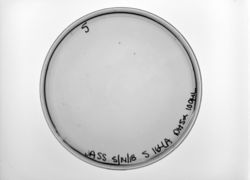
[[Image:JASS S164A 052018.jpg|thumb|left|250px|Figure 1: Bacterial Transformation]] | [[Image:JASS S164A 052018.jpg|thumb|left|250px|Figure 1: Bacterial Transformation]] | ||
| - | To add an image of your experiments, you first need to upload the image. | + | To add an image of your experiments, you first need to upload the image. |
| + | To see how to embed the image as I've shown above, use the following code: | ||
| - | + | Image:[insert filename.jpg]|thumb|left|250px|Figure [insert figure#]: [insert your caption] | |
| + | You can resize your image, or set the location to right or center as you wish. | ||
== Background == | == Background == | ||
Revision as of 17:02, 16 May 2018
Contents |
Transcription Factor AphB from V. cholerae
To add an image of your experiments, you first need to upload the image.
To see how to embed the image as I've shown above, use the following code:
Image:[insert filename.jpg]|thumb|left|250px|Figure [insert figure#]: [insert your caption]
You can resize your image, or set the location to right or center as you wish.
Background
Solution Studies
Structural highlights
Each AphB comprises an N-terminal (DBD) and a C-terminal (RD). The DBD is connected to the RD via the . AphB subunits are found in two distinct conformations within the tetramer, as determined by the angle between the linker helix and the RD. In the form, the RD and linker helix form an angle of 85 degrees, whereas this angle is 150 degrees in the extended form. Interestingly, the LH forms the dimerization interface between one compact and one extended subunit.
The tetramer (chains A, B, C and D) can be described as a dimer of dimers. Each of two identical dimers are composed of a compact (A and C) and extended (B and D) monomer. The compact and extended subunits differ from one another in the angle between the linker helix and the RD, which is 85° in the compact subunits and 150° in the extended subunits (Fig. 1A). The conformational variability in the AphB subunits can be attributed to the flexible loop connecting the linker helix to the regulatory domain.
The tetramer assembles via two distinct dimerization interfaces (Fig. 2A). The N‐terminal dimerization interface (1142 Å2) is primarily made up of an antiparallel coiled‐coil of the two linker helices, held together by hydrophobic, polar and ionic interactions (Fig. 2A, left inset). The C‐terminal dimerization interface (1414 Å2) forms between regulatory domains, which pack against each other in a head‐to‐tail manner (Fig. 2A, right inset). To form the AphB tetramer, each compact subunit interacts with the regulatory domain of one extended subunit and with the linker helix of the other extended subunit (Fig. 2A and B).
</StructureSection>

