User:Ashley Crotteau/Sandbox1
From Proteopedia
(Difference between revisions)
| Line 16: | Line 16: | ||
The ΔSET7/9 consists of the SET domain along with the pre- and post-SET regions.<ref name="Schubert" /><ref name="Yeates" /> The pre- and post-SET regions are adjacent to SET domain and are cysteine rich.<ref name="Schubert" /><ref name="Yeates" /> The pre-SET cysteine region is located near the N-terminal where the post-SET region is located near the C-terminal of the domain.<ref name="Schubert" /><ref name="Yeates" /> These regions are said to play an important role in substrate recognition and enzymatic activity.<ref name="Schubert" /><ref name="Yeates" /> The cysteine regions are not shown in the crystal structure as only residues 117-366 were crystalized.<ref name="Xiao" /> | The ΔSET7/9 consists of the SET domain along with the pre- and post-SET regions.<ref name="Schubert" /><ref name="Yeates" /> The pre- and post-SET regions are adjacent to SET domain and are cysteine rich.<ref name="Schubert" /><ref name="Yeates" /> The pre-SET cysteine region is located near the N-terminal where the post-SET region is located near the C-terminal of the domain.<ref name="Schubert" /><ref name="Yeates" /> These regions are said to play an important role in substrate recognition and enzymatic activity.<ref name="Schubert" /><ref name="Yeates" /> The cysteine regions are not shown in the crystal structure as only residues 117-366 were crystalized.<ref name="Xiao" /> | ||
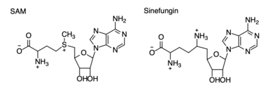
| - | The SET domain is mostly defined by | + | The SET domain is mostly defined by turns and loops, which connect secondary structures, with the few <scene name='81/811707/Beta_sheets/3'>antiparallel β-sheets</scene>.<ref name="Schubert" /> <scene name='81/811707/Beta-hairpin/3'>Residues 337-349</scene> form a β-hairpin that sticks out at a right angle to the surface of the enzyme.<ref name="Xiao" /> The following three residues (<scene name='81/811707/Sharp_bend/3'>350-352</scene>) accommodate a sharp bend in the peptide chain and the end of the protein takes on an <scene name='81/811707/C-term_alpha_helix/2'>α-helical conformation</scene>.<ref name="Xiao" /> The two most defining features of the SET domain are the C-terminal tyrosine and the <scene name='81/811707/Variable_knot/3'>knot-like fold</scene>. These two components have been recognized to be essential for S-adenosyl-L-methionine(SAM) binding and catalysis, which is shown as <scene name='81/811707/Sam_isolated/2'>S-adenosyl-L-homocysteine</scene>(SAH) in the structure after methylation of the histone.<ref name="Schubert" /> <ref name="Yeates" /> <ref name="Huang" /> The knot-like fold contains the binding sites for the cofactor SAM and the peptide substrate.<ref name="Licciardello" /> |
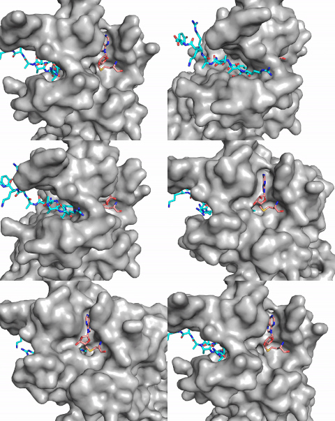
[[Image:Methyl1.gif|300 px|right|thumb|Figure 1: Image of substrate bound on one side of SET7/9 (PDB 1o9s) with the lysine target in the active site channel (cyan) and S-adenosyl homocysteine (pink) bound on the opposite face of the enzyme]] | [[Image:Methyl1.gif|300 px|right|thumb|Figure 1: Image of substrate bound on one side of SET7/9 (PDB 1o9s) with the lysine target in the active site channel (cyan) and S-adenosyl homocysteine (pink) bound on the opposite face of the enzyme]] | ||
Revision as of 20:53, 24 April 2019
H. sapiens Lysine Methyltransferase, SET 7/9
| |||||||||||
References
[3] [8] [5] [6] [7] [9] [10] [11] [1] [2] [4]
- ↑ 1.0 1.1 DesJarlais R, Tummino PJ. Role of Histone-Modifying Enzymes and Their Complexes in Regulation of Chromatin Biology. Biochemistry. 2016 Mar 22;55(11):1584-99. doi: 10.1021/acs.biochem.5b01210. Epub , 2016 Jan 26. PMID:26745824 doi:http://dx.doi.org/10.1021/acs.biochem.5b01210
- ↑ 2.0 2.1 Marino-Ramirez L, Kann MG, Shoemaker BA, Landsman D. Histone structure and nucleosome stability. Expert Rev Proteomics. 2005 Oct;2(5):719-29. PMID:16209651 doi:http://dx.doi.org/10.1586/14789450.2.5.719
- ↑ 3.00 3.01 3.02 3.03 3.04 3.05 3.06 3.07 3.08 3.09 3.10 Xiao B, Jing C, Wilson JR, Walker PA, Vasisht N, Kelly G, Howell S, Taylor IA, Blackburn GM, Gamblin SJ. Structure and catalytic mechanism of the human histone methyltransferase SET7/9. Nature. 2003 Feb 6;421(6923):652-6. Epub 2003 Jan 22. PMID:12540855 doi:10.1038/nature01378
- ↑ 4.0 4.1 4.2 doi: https://dx.doi.org/10.15406/mojcsr.2016.03.00047
- ↑ 5.0 5.1 5.2 5.3 5.4 5.5 5.6 Schubert HL, Blumenthal RM, Cheng X. Many paths to methyltransfer: a chronicle of convergence. Trends Biochem Sci. 2003 Jun;28(6):329-35. PMID:12826405
- ↑ 6.0 6.1 6.2 6.3 6.4 6.5 Yeates TO. Structures of SET domain proteins: protein lysine methyltransferases make their mark. Cell. 2002 Oct 4;111(1):5-7. PMID:12372294
- ↑ 7.0 7.1 Huang S, Shao G, Liu L. The PR domain of the Rb-binding zinc finger protein RIZ1 is a protein binding interface and is related to the SET domain functioning in chromatin-mediated gene expression. J Biol Chem. 1998 Jun 26;273(26):15933-9. PMID:9632640
- ↑ 8.0 8.1 doi: https://dx.doi.org/10.1016/C2014-0-02189-2
- ↑ 9.0 9.1 9.2 9.3 9.4 9.5 Del Rizzo PA, Couture JF, Dirk LM, Strunk BS, Roiko MS, Brunzelle JS, Houtz RL, Trievel RC. SET7/9 catalytic mutants reveal the role of active site water molecules in lysine multiple methylation. J Biol Chem. 2010 Oct 8;285(41):31849-58. Epub 2010 Aug 1. PMID:20675860 doi:http://dx.doi.org/10.1074/jbc.M110.114587
- ↑ 10.0 10.1 10.2 10.3 10.4 Sun G, Reddy MA, Yuan H, Lanting L, Kato M, Natarajan R. Epigenetic histone methylation modulates fibrotic gene expression. J Am Soc Nephrol. 2010 Dec;21(12):2069-80. doi: 10.1681/ASN.2010060633. Epub 2010, Oct 7. PMID:20930066 doi:http://dx.doi.org/10.1681/ASN.2010060633
- ↑ 11.0 11.1 Tian X, Zhang S, Liu HM, Zhang YB, Blair CA, Mercola D, Sassone-Corsi P, Zi X. Histone lysine-specific methyltransferases and demethylases in carcinogenesis: new targets for cancer therapy and prevention. Curr Cancer Drug Targets. 2013 Jun;13(5):558-79. doi:, 10.2174/1568009611313050007. PMID:23713993 doi:http://dx.doi.org/10.2174/1568009611313050007
Student Contributors
Ashley Crotteau
Parker Hiday
Lauren Allman


