This old version of Proteopedia is provided for student assignments while the new version is undergoing repairs. Content and edits done in this old version of Proteopedia after March 1, 2026 will eventually be lost when it is retired in about June of 2026.
Apply for new accounts at the new Proteopedia. Your logins will work in both the old and new versions.
Methyl CpG binding protein
From Proteopedia
(Difference between revisions)
| Line 17: | Line 17: | ||
== Structural highlights == | == Structural highlights == | ||
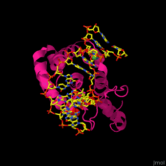
| - | <scene name='59/597003/Cv/ | + | <scene name='59/597003/Cv/3'>Thymine from the mismatched G:T pair flips out from the DNA strand and interchelates with MBD4 residues Lys and Leu</scene><ref>PMID:22740654</ref>. |
</StructureSection> | </StructureSection> | ||
Revision as of 13:45, 11 July 2019
| |||||||||||
3D structures of methyl CpG binding protein
Updated on 11-July-2019
References
- ↑ Wade PA. Methyl CpG-binding proteins and transcriptional repression. Bioessays. 2001 Dec;23(12):1131-7. PMID:11746232 doi:http://dx.doi.org/10.1002/bies.10008
- ↑ Van den Veyver IB, Zoghbi HY. Mutations in the gene encoding methyl-CpG-binding protein 2 cause Rett syndrome. Brain Dev. 2001 Dec;23 Suppl 1:S147-51. PMID:11738862
- ↑ Hashimoto H, Zhang X, Cheng X. Excision of thymine and 5-hydroxymethyluracil by the MBD4 DNA glycosylase domain: structural basis and implications for active DNA demethylation. Nucleic Acids Res. 2012 Jun 27. PMID:22740654 doi:10.1093/nar/gks628
