Methyl CpG Binding Protein 2
From Proteopedia
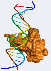
Methyl CpG Binding Protein 2 (MeCP2) is a transcriptional repressor that plays a critical gene silencing role. It binds the methylated BDNF DNA sequence with exquisite specificity and recruits repressor complexes which include chromatin-remodeling enzymes.[1] MeCP2, encoded by an X-linked gene, has been found in most tissues but is primarily localized to cortical neurons where it is believed to help modulate neuronal maturity and plasticity.[2] It also plays a crucial role in synaptogenesis and in maintaining proper neuronal function.[1] Of particular note, mutations within the MeCP2 gene are responsible for over 95% of cases of Rett Syndrome. Rett Syndrome (RTT) is an autism spectrum neurodevelopmental disorder caused by the expression of mutant MeCP2 . RTT patients present with abnormal neuronal morphology and have a large spectrum of autistic characteristics such as impaired motor function and impaired developmental skills. These symptoms become first evident 6-18 months after birth.[3] Intriguingly, Guy et al. demonstrated that the restoration of neuronal function by knock-in expression of MeCP2 in mice can reverse many of the RTT phenotypic traits.[4] MeCP2 is present in mature nerve cells and is involved in turning off several genes. Structure of MeCP2The crystal structure of the DNA binding domain of MeCP2 was solved by Ho et al. in 2008 providing researchers with critical new information about the cause of RTT at the molecular level. The MeCP2 DNA Binding Domain (MBD) binds the methylated BDNF DNA sequence using a predominantly hydrophilic pocket. MeCP2 recognition of the mCpG DNA sequence involves five water molecules each making unique CH-O hydrogen bonds. water-22 forms hydrogen bonds with Asp 121, water-24, water-21, and N4 of m5C33. water-24 forms hydrogen bonds with Tyr 123, Arg 133, water 22, as well as m5C8 and a CH-O hydrogen bond interaction with the methyl group of m5C8. The only residues that directly interact with DNA bases are Asp 121, which forms hydrogen bond with the methyl group of m5C8, and Arg 111, and Arg 133, each of which form hydrogen bonds with a guanine DNA base. Both of these “Arginine Fingers” lie in the same plane as the guanines and are secured into this position by salt bridges with Asp 121 and Glu 137. These interactions place the gaunidines directly above/below the methyl groups of the methyl-cytidines.[1] The C-terminal region of the MBD includes an unusual tandem ASX-ST motif. This motif consists of an ASX turn (Residues 156-158) directly followed by an ST motif (158-161). The ASX turn structure is stabilized by a hydrogen bond interaction connecting the nitrogen of Thr 158 and Asp 156. The ST motif is stabilized by hydrogen bonds to Thr 158. T158M, which is the most common missense mutation causing Rett Syndrome abolishes DNA binding because it disrupts this ASX-ST motif. Another well-known RTT inducing mutation, R106W, disrupts the ASX-ST motif stabilizing hydrogen bonds between Arg 106 and Thr 158 and Val 159. This ASX-ST motif stabilizes MeCP2’s interaction with DNA by specifically binding to the AATT minor groove which is 3 angstroms narrower than a typical DNA minor groove, due to the consecutive A/T bases. This unique trait helps account for MeCP2’s exquisite precision.[1] Overall, the solved crystal structure of the MBD of MeCP2 reveals why certain known mutations are so deleterious as to cause Rett Syndrome and hopefully elucidates potential avenues for pharmaceutical intervention. See also Neurodevelopmental Disorders.
|
|
Contents |
Page Development
This article was developed based on lectures given in Chemistry 543 by Prof. Clarence E. Schutt at Princeton University.
3D structures of MeCP2
See Methyl CpG binding protein.
See Also
- Introduction to Evolutionary Conservation which uses MeCP2 as an example.
- Brief introduction to Rett Syndrome
References
- ↑ 1.0 1.1 1.2 1.3 Ho KL, McNae IW, Schmiedeberg L, Klose RJ, Bird AP, Walkinshaw MD. MeCP2 binding to DNA depends upon hydration at methyl-CpG. Mol Cell. 2008 Feb 29;29(4):525-31. PMID:18313390 doi:10.1016/j.molcel.2007.12.028
- ↑ Zoghbi HY. Postnatal neurodevelopmental disorders: meeting at the synapse? Science. 2003 Oct 31;302(5646):826-30. PMID:14593168 doi:10.1126/science.1089071
- ↑ Marchetto MC, Carromeu C, Acab A, Yu D, Yeo GW, Mu Y, Chen G, Gage FH, Muotri AR. A model for neural development and treatment of Rett syndrome using human induced pluripotent stem cells. Cell. 2010 Nov 12;143(4):527-39. PMID:21074045 doi:10.1016/j.cell.2010.10.016
- ↑ Guy J, Gan J, Selfridge J, Cobb S, Bird A. Reversal of neurological defects in a mouse model of Rett syndrome. Science. 2007 Feb 23;315(5815):1143-7. Epub 2007 Feb 8. PMID:17289941 doi:10.1126/science.1138389
Proteopedia Page Contributors and Editors (what is this?)
David Canner, Michal Harel, Eric Martz, Jaime Prilusky, Alexander Berchansky