We apologize for Proteopedia being slow to respond. For the past two years, a new implementation of Proteopedia has been being built. Soon, it will replace this 18-year old system. All existing content will be moved to the new system at a date that will be announced here.
Sandbox Reserved 1111
From Proteopedia
(Difference between revisions)
| Line 6: | Line 6: | ||
== Generalities == | == Generalities == | ||
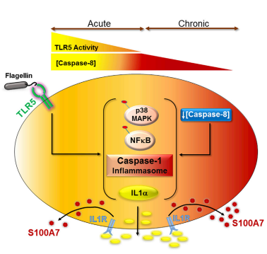
| - | The structure <scene name='82/829364/1psr/1'>1PSR</scene> is found in the human psoriasin, also called [https://en.wikipedia.org/wiki/S100A7 S100A7]. This protein belongs to the family of [http://proteopedia.org/wiki/index.php/Psoriasin S100] proteins. They are called like this because of their solubility in a saturated solution with ammonium sulfate at neutral pH <ref> Sedaghat F, Notopoulos A (October 2008). "S100 protein family and its application in clinical practice". ''Hippokratia''. '''4''': 198-204. https://www.ncbi.nlm.nih.gov/pmc/articles/PMC2580040/pdf/hippokratia-12-198.pdf </ref>. It is a family of 21 proteins of low molecular weights with a high frequence of homologous sequence (often between 40% and 50%). Those proteins are found in cells as homo and heterodimers. Each monomer have a similar structure, beginning with the N-terminal EF Hand and the four-turn <scene name='82/829364/Helix_1/2'>Helix I</scene> which leads to a <scene name='82/829364/Loop/1'>loop</scene>. Then, there is the three-turn <scene name='82/829364/Helix_ii/1'>Helix II</scene>, the two-turn <scene name='82/829364/Helix_iii/1'>Helix III</scene> and finally the five-turn <scene name='82/829364/Helix_iv/1'>Helix IV</scene>. One of their main properties is their ability to bind the calcium. They share some common structures such as two helix-loop-helix structures which are calcium-binding domains more specifically the loop from <scene name='82/829364/Calcium_binding_site/1'>Asp62 to Asp70</scene>. All the S100 proteins have different functions in many various cell types. They have significant roles in calcium-associated signal transduction. They play the roles of calcium sensors proteins that regulate the function or distribution of specific target proteins <ref>Eckert R, Broome AM, Ruse M, Robinson N, Ryan D, Lee K (July 2004). "S100 Proteins in the Epidermis". ''Journal of Investigative Dermatology''. '''123'''(1): 23-33.https://doi.org/10.1111/j.0022-202X.2004.22719.x</ref>. | + | The structure <scene name='82/829364/1psr/1'>1PSR</scene> is found in the human psoriasin, also called [https://en.wikipedia.org/wiki/S100A7 S100A7]. This protein belongs to the family of [http://proteopedia.org/wiki/index.php/Psoriasin S100] proteins whitch are involed in the skin celle division and differenciation. They are called like this because of their solubility in a saturated solution with ammonium sulfate at neutral pH <ref> Sedaghat F, Notopoulos A (October 2008). "S100 protein family and its application in clinical practice". ''Hippokratia''. '''4''': 198-204. https://www.ncbi.nlm.nih.gov/pmc/articles/PMC2580040/pdf/hippokratia-12-198.pdf </ref>. It is a family of 21 proteins of low molecular weights with a high frequence of homologous sequence (often between 40% and 50%). Those proteins are found in cells as homo and heterodimers. Each monomer have a similar structure, beginning with the N-terminal EF Hand and the four-turn <scene name='82/829364/Helix_1/2'>Helix I</scene> which leads to a <scene name='82/829364/Loop/1'>loop</scene>. Then, there is the three-turn <scene name='82/829364/Helix_ii/1'>Helix II</scene>, the two-turn <scene name='82/829364/Helix_iii/1'>Helix III</scene> and finally the five-turn <scene name='82/829364/Helix_iv/1'>Helix IV</scene>. One of their main properties is their ability to bind the calcium. They share some common structures such as two helix-loop-helix structures which are calcium-binding domains more specifically the loop from <scene name='82/829364/Calcium_binding_site/1'>Asp62 to Asp70</scene>. All the S100 proteins have different functions in many various cell types. They have significant roles in calcium-associated signal transduction. They play the roles of calcium sensors proteins that regulate the function or distribution of specific target proteins <ref>Eckert R, Broome AM, Ruse M, Robinson N, Ryan D, Lee K (July 2004). "S100 Proteins in the Epidermis". ''Journal of Investigative Dermatology''. '''123'''(1): 23-33.https://doi.org/10.1111/j.0022-202X.2004.22719.x</ref>. |
== Human Psoriasin == | == Human Psoriasin == | ||
Revision as of 21:40, 17 January 2020
| This Sandbox is Reserved from 25/11/2019, through 30/9/2020 for use in the course "Structural Biology" taught by Bruno Kieffer at the University of Strasbourg, ESBS. This reservation includes Sandbox Reserved 1091 through Sandbox Reserved 1115. |
To get started:
More help: Help:Editing |
1 PSR
| |||||||||||
References
- ↑ Sedaghat F, Notopoulos A (October 2008). "S100 protein family and its application in clinical practice". Hippokratia. 4: 198-204. https://www.ncbi.nlm.nih.gov/pmc/articles/PMC2580040/pdf/hippokratia-12-198.pdf
- ↑ Eckert R, Broome AM, Ruse M, Robinson N, Ryan D, Lee K (July 2004). "S100 Proteins in the Epidermis". Journal of Investigative Dermatology. 123(1): 23-33.https://doi.org/10.1111/j.0022-202X.2004.22719.x
- ↑ Hagens G, Masouyé I, Augsburger E, Hotz R, Saurat JH, Siegenthaler G (April 1999) "Calcium-binding protein S100A7 and epidermal-type fatty acid-binding protein are associated in the cytosol of human keratinocytes" Biochem J. 339(2): 419-427: https://www.ncbi.nlm.nih.gov/pmc/articles/PMC1220173/
- ↑ Chmurzyńska A (March 2006). "The multigene family of fatty acid-binding proteins (FABPs): Function, structure and polymorphism".Journal of Applied Genetics.47(1): 39-48.https://link.springer.com/article/10.1007/BF03194597
- ↑ Brodersen DE, Etzerodt M, Madsen P, Celis JE, Thøgersen HC, Nyborg J, Kjeldgaard M (April 1998). "EF-hands at atomic resolution: the structure of human psoriasin(S100A7) solved by MAD phasing". Structure.6: 477-489.https://www.cell.com/structure/pdf/S0969-2126(98)00049-5.pdf
- ↑ Murray J, Boulanger M (April 2010). "S100A7 (S100 calcium binding protein A7)". Atlas of Genetics and Cytogeneticsin Oncology and Haematology. 15(1): 59-64. : http://AtlasGeneticsOncology.org/Genes/S100A7ID42194ch1q21.html
- ↑ S Alowami, G Qing, E Emberley, L Snell & PH Watson (2003). "Psoriasin , (S100A7) expression is altered during skin tumorigenesis" BMC Dermatology.3(1):
- ↑ AK Ekman, J Vegfors, C Bivik Eding, C Enerbäck (December 2016). "Overexpression of Psoriasin (S100A7) Contributes to Dysregulated Differentiation in Psoriasis", Acta Derm Venereol. 97: https://www.medicaljournals.se/acta/content/html/10.2340/00015555-2596
- ↑ KC Lee , RL Eckert (April 2007). "S100A7 (Psoriasin) – Mechanism of Antibacterial Action in Wounds", Journal of Investigative Dermatology 127(4): 945-957 https://www.sciencedirect.com/science/article/pii/S0022202X15333145
- ↑ T Bhatt, A Bhosale, B Bajantri, MS Mathapathi, A Rizvi, G Scita, A Majumdar and C Jamora (November 2019) "Sustained Secretion of the Antimicrobial Peptide S100A7 Is Dependent on the Downregulation of Caspase-8", Cell Reports. 29(9): 2546-2555 https://doi.org/10.1016/j.celrep.2019.10.090

