Journal:Acta Cryst D:S205979832000501X
From Proteopedia
(Difference between revisions)

| Line 17: | Line 17: | ||
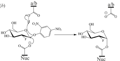
[[Image:Fig7b.png|left|400px|thumb|'''Figure 7''' (b) Mechanism of 2F-DNPGlc hydrolysis by GBA to generate the covalent glycosyl-enzyme intermediate.]] | [[Image:Fig7b.png|left|400px|thumb|'''Figure 7''' (b) Mechanism of 2F-DNPGlc hydrolysis by GBA to generate the covalent glycosyl-enzyme intermediate.]] | ||
| - | <scene name='84/842878/Cv/9'>Crystal structure of GBA monomer obtained at 0.98 Å resolution</scene> (PDB [[6tn1]]). Domain I (residues 1-27 and 383-414) in orange, domain II (residues 30-75 and 431-497) in violet and domain III (residues 76–381 and 416-430) in blue. ''N''-glycans are shown in ball-and-stick representation. <scene name='84/842878/Cv/11'>Overlay of GBA unliganded structure with BTP complexed structure</scene> (PDB [[6tjk]]). Red indicates areas of high RMSD between the protein backbones. Loop 1 contains residues 27-31, loop 2 comprises residues 314-319 and loop 3 contains residues 344-350. <scene name='84/842878/Cv/13'>Active site of GBA unliganded crystal structure (blue) overlaid with active site residues of the BTP complex structure (gold) (PDB 6TJK) and Cerezyme (green) (PDB 6TJJ)</scene>. A magnesium ion (peach) coordinated by four waters (grey), Glu340 (nuc) and Glu235 (a/b), occupies the active site. | + | <scene name='84/842878/Cv/9'>Crystal structure of GBA monomer obtained at 0.98 Å resolution</scene> (PDB [[6tn1]]). Domain I (residues 1-27 and 383-414) in orange, domain II (residues 30-75 and 431-497) in violet and domain III (residues 76–381 and 416-430) in blue. ''N''-glycans are shown in ball-and-stick representation. <scene name='84/842878/Cv/11'>Overlay of GBA unliganded structure with bis-Tris propane (BTP) complexed structure</scene> (PDB [[6tjk]]). Red indicates areas of high RMSD between the protein backbones. Loop 1 contains residues 27-31, loop 2 comprises residues 314-319 and loop 3 contains residues 344-350. <scene name='84/842878/Cv/13'>Active site of GBA unliganded crystal structure (blue) overlaid with active site residues of the BTP complex structure (gold) (PDB 6TJK) and Cerezyme (green) (PDB 6TJJ)</scene>. A magnesium ion (peach) coordinated by four waters (grey), Glu340 (nuc) and Glu235 (a/b), occupies the active site. |
<b>References</b><br> | <b>References</b><br> | ||
Revision as of 18:43, 27 June 2021
| |||||||||||
Proteopedia Page Contributors and Editors (what is this?)
This page complements a publication in scientific journals and is one of the Proteopedia's Interactive 3D Complement pages. For aditional details please see I3DC.


