This old version of Proteopedia is provided for student assignments while the new version is undergoing repairs. Content and edits done in this old version of Proteopedia after March 1, 2026 will eventually be lost when it is retired in about June of 2026.
Apply for new accounts at the new Proteopedia. Your logins will work in both the old and new versions.
1th6
From Proteopedia
(Difference between revisions)
| Line 3: | Line 3: | ||
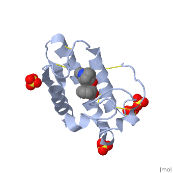
<StructureSection load='1th6' size='340' side='right'caption='[[1th6]], [[Resolution|resolution]] 1.23Å' scene=''> | <StructureSection load='1th6' size='340' side='right'caption='[[1th6]], [[Resolution|resolution]] 1.23Å' scene=''> | ||
== Structural highlights == | == Structural highlights == | ||
| - | <table><tr><td colspan='2'>[[1th6]] is a 1 chain structure with sequence from [ | + | <table><tr><td colspan='2'>[[1th6]] is a 1 chain structure with sequence from [https://en.wikipedia.org/wiki/Daboia_russellii_russellii Daboia russellii russellii]. Full crystallographic information is available from [http://oca.weizmann.ac.il/oca-bin/ocashort?id=1TH6 OCA]. For a <b>guided tour on the structure components</b> use [https://proteopedia.org/fgij/fg.htm?mol=1TH6 FirstGlance]. <br> |
| - | </td></tr><tr id='ligand'><td class="sblockLbl"><b>[[Ligand|Ligands:]]</b></td><td class="sblockDat"><scene name='pdbligand=OIN:(1R,5S)-8-METHYL-8-AZABICYCLO[3.2.1]OCT-3-YL+(2R)-3-HYDROXY-2-PHENYLPROPANOATE'>OIN</scene>, <scene name='pdbligand=SO4:SULFATE+ION'>SO4</scene></td></tr> | + | </td></tr><tr id='ligand'><td class="sblockLbl"><b>[[Ligand|Ligands:]]</b></td><td class="sblockDat" id="ligandDat"><scene name='pdbligand=OIN:(1R,5S)-8-METHYL-8-AZABICYCLO[3.2.1]OCT-3-YL+(2R)-3-HYDROXY-2-PHENYLPROPANOATE'>OIN</scene>, <scene name='pdbligand=SO4:SULFATE+ION'>SO4</scene></td></tr> |
| - | <tr id='related'><td class="sblockLbl"><b>[[Related_structure|Related:]]</b></td><td class="sblockDat">[[1q7a|1q7a]], [[1sv3|1sv3]], [[1tg1|1tg1]], [[1tg4|1tg4]], [[1tgm|1tgm]], [[1ti0|1ti0]]</td></tr> | + | <tr id='related'><td class="sblockLbl"><b>[[Related_structure|Related:]]</b></td><td class="sblockDat"><div style='overflow: auto; max-height: 3em;'>[[1q7a|1q7a]], [[1sv3|1sv3]], [[1tg1|1tg1]], [[1tg4|1tg4]], [[1tgm|1tgm]], [[1ti0|1ti0]]</div></td></tr> |
| - | <tr id='activity'><td class="sblockLbl"><b>Activity:</b></td><td class="sblockDat"><span class='plainlinks'>[ | + | <tr id='activity'><td class="sblockLbl"><b>Activity:</b></td><td class="sblockDat"><span class='plainlinks'>[https://en.wikipedia.org/wiki/Phospholipase_A(2) Phospholipase A(2)], with EC number [https://www.brenda-enzymes.info/php/result_flat.php4?ecno=3.1.1.4 3.1.1.4] </span></td></tr> |
| - | <tr id='resources'><td class="sblockLbl"><b>Resources:</b></td><td class="sblockDat"><span class='plainlinks'>[ | + | <tr id='resources'><td class="sblockLbl"><b>Resources:</b></td><td class="sblockDat"><span class='plainlinks'>[https://proteopedia.org/fgij/fg.htm?mol=1th6 FirstGlance], [http://oca.weizmann.ac.il/oca-bin/ocaids?id=1th6 OCA], [https://pdbe.org/1th6 PDBe], [https://www.rcsb.org/pdb/explore.do?structureId=1th6 RCSB], [https://www.ebi.ac.uk/pdbsum/1th6 PDBsum], [https://prosat.h-its.org/prosat/prosatexe?pdbcode=1th6 ProSAT]</span></td></tr> |
</table> | </table> | ||
== Function == | == Function == | ||
| - | [[ | + | [[https://www.uniprot.org/uniprot/PA2B8_DABRR PA2B8_DABRR]] Snake venom phospholipase A2 (PLA2) that shows weak neurotoxicity and medium anticoagulant effects by binding to factor Xa (F10) and inhibiting the prothrombinase activity (IC(50) is 130 nM) (PubMed:18062812). It also damages vital organs such as lung, liver and kidney, displays edema-inducing activities when injected into the foot pads of mice and induces necrosis of muscle cells when injected into the thigh muscle. Has a low enzymatic activity. PLA2 catalyzes the calcium-dependent hydrolysis of the 2-acyl groups in 3-sn-phosphoglycerides.<ref>PMID:18062812</ref> <ref>PMID:2115497</ref> <ref>PMID:8835338</ref> |
== Evolutionary Conservation == | == Evolutionary Conservation == | ||
[[Image:Consurf_key_small.gif|200px|right]] | [[Image:Consurf_key_small.gif|200px|right]] | ||
| Line 23: | Line 23: | ||
==See Also== | ==See Also== | ||
| - | *[[Atropine|Atropine]] | ||
*[[Phospholipase A2|Phospholipase A2]] | *[[Phospholipase A2|Phospholipase A2]] | ||
*[[Phospholipase A2 3D structures|Phospholipase A2 3D structures]] | *[[Phospholipase A2 3D structures|Phospholipase A2 3D structures]] | ||
Revision as of 09:27, 29 September 2021
Crystal structure of phospholipase A2 in complex with atropine at 1.23A resolution
| |||||||||||