Sandbox Reserved 1694
From Proteopedia
(Difference between revisions)
| Line 23: | Line 23: | ||
== Structural highlights == | == Structural highlights == | ||
[[Image:h_bonds_2.png | thumb]] | [[Image:h_bonds_2.png | thumb]] | ||
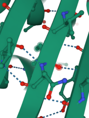
| - | INPP1D54A contains 13 | + | INPP1D54A contains 13 <scene name='89/892737/Secondary_structures/5'>secondary strucutures</scene> of which 10 are alpha helices and 3 are beta-sheets. Percentage-wise, this enzyme is 77% alpha-helices, 23% beta-sheets. Alpha helix 8 and beta-sheet 2 each contain one catalytic amino acid. Also, helix 6 and 7 and beta-sheet 2 form important interactions with the substrate (have 6 of the 9 amino acids that interact with the substrate). These alpha-helices and two large anti-parallel beta-sheets help determine the structure and function of this protein. Also, they provide stabilization through hydrogen bonding (shown in the picture on the right) that solidifies the conformation of beta-sheets and helices. This protein is already folded in its <scene name='89/892737/Tert_structure/2'>tertiary structure</scene> which is colored coated to show polar (pink) and hydrophobic (grey) amino acids indicating potential intermolecular interactions such as hydrophobic interaction, disulfide bridge, ionic, and hydrogen bonds. In addition, INPP1D54A and INPP1 do not exhibit a quaternary structure since it only consists of one subunit rather than two or more subunits. |
A <scene name='89/892737/Space_fill/4'>space fill</scene> molecular model of INPP1D54A displays how much space the atoms within the protein occupy and gives the relative dimensions and shape of the protein. It also shows some of the <scene name='89/892737/Space_fill_2/2'>water molecules</scene> (red) that interact with the protein and play an important part in the nucleophilic attack on the phosphate group removed from as the substrate binds to the active site. This image with the substrate transparent gives insight into the pocket where the substrate binds to the active site on the enzyme. | A <scene name='89/892737/Space_fill/4'>space fill</scene> molecular model of INPP1D54A displays how much space the atoms within the protein occupy and gives the relative dimensions and shape of the protein. It also shows some of the <scene name='89/892737/Space_fill_2/2'>water molecules</scene> (red) that interact with the protein and play an important part in the nucleophilic attack on the phosphate group removed from as the substrate binds to the active site. This image with the substrate transparent gives insight into the pocket where the substrate binds to the active site on the enzyme. | ||
Current revision
| This Sandbox is Reserved from 10/01/2021 through 01/01//2022 for use in Biochemistry taught by Bonnie Hall at Grand View University, Des Moines, USA. This reservation includes Sandbox Reserved 1690 through Sandbox Reserved 1699. |
To get started:
More help: Help:Editing |
Inositol polyphosphate 1-Phosphatase (INPP1) D54A
| |||||||||||
References
- ↑ Hanson, R. M., Prilusky, J., Renjian, Z., Nakane, T. and Sussman, J. L. (2013), JSmol and the Next-Generation Web-Based Representation of 3D Molecular Structure as Applied to Proteopedia. Isr. J. Chem., 53:207-216. doi:http://dx.doi.org/10.1002/ijch.201300024
- ↑ Herraez A. Biomolecules in the computer: Jmol to the rescue. Biochem Mol Biol Educ. 2006 Jul;34(4):255-61. doi: 10.1002/bmb.2006.494034042644. PMID:21638687 doi:10.1002/bmb.2006.494034042644
- ↑ 3.0 3.1 3.2 Dollins DE, Xiong JP, Endo-Streeter S, Anderson DE, Bansal VS, Ponder JW, Ren Y, York JD. A Structural Basis for Lithium and Substrate Binding of an Inositide Phosphatase. J Biol Chem. 2020 Nov 10. pii: RA120.014057. doi: 10.1074/jbc.RA120.014057. PMID:33172890 doi:http://dx.doi.org/10.1074/jbc.RA120.014057


