We apologize for Proteopedia being slow to respond. For the past two years, a new implementation of Proteopedia has been being built. Soon, it will replace this 18-year old system. All existing content will be moved to the new system at a date that will be announced here.
Sandbox Reserved 1653
From Proteopedia
(Difference between revisions)
| Line 99: | Line 99: | ||
Because Piezo1 is implicated in the functioning of many cells and organs, modifications on its structure lead to diseases. | Because Piezo1 is implicated in the functioning of many cells and organs, modifications on its structure lead to diseases. | ||
| - | For instance, Hereditary Xerocytosis (HX) is a rare disease, also called [https://en.wikipedia.org/wiki/Hereditary_stomatocytosis Dehydrated hereditary stomatocytosis] (DHS). This genetic disease leads to impaired red blood cell (RBC) membrane properties that affect intracellular cation concentrations.<ref name="Dehydrated"> DOI 10.1038/ncomms2899</ref> The RBCs are abnormally shaped and they result in [https://en.wikipedia.org/wiki/Hemolytic_anemia haemolytic anaemia]. Those modifications are due to mutations on the FAM38A gene on chromosome 16 which encodes for Piezo 1. Piezo1 is expressed in the plasma membranes of RBCs, and its role is to control RBCs’ osmolarity. It also plays a prevalent role in the [https://en.wikipedia.org/wiki/Erythropoiesis erythroid differentiation]. Mutations in Piezo1 distort mechanosensitive channel regulation, leading to increased cation transport in erythroid cells. Those mutations affect different parts of the channel. For instance, six gain-of-function mutations, gathered in the central core region of the Piezo channel structure, are directly linked to the decrease of inactivation rate of the channel. Mutations in the N-terminal part of the protein also have a role in channel gating. Therefore, not every channel is affected in the same way and by the same mutation. Indeed it depends on the environment, the permeability of RBC and the combination of mutations. | + | For instance, Hereditary Xerocytosis (HX) is a rare disease, also called [https://en.wikipedia.org/wiki/Hereditary_stomatocytosis Dehydrated hereditary stomatocytosis] (DHS). This genetic disease leads to impaired red blood cell (RBC) membrane properties that affect intracellular cation concentrations.<ref name="Dehydrated"> DOI 10.1038/ncomms2899</ref> The RBCs are abnormally shaped and they result in [https://en.wikipedia.org/wiki/Hemolytic_anemia haemolytic anaemia]. Those modifications are due to mutations on the FAM38A gene on chromosome 16 which encodes for Piezo 1. Piezo1 is expressed in the plasma membranes of RBCs, and its role is to control RBCs’ osmolarity. It also plays a prevalent role in the [https://en.wikipedia.org/wiki/Erythropoiesis erythroid differentiation]. Mutations in Piezo1 distort mechanosensitive channel regulation, leading to increased cation transport in erythroid cells. Those mutations affect different parts of the channel. For instance, six gain-of-function mutations, gathered in the central core region of the Piezo channel structure, are directly linked to the decrease of inactivation rate of the channel. Mutations in the N-terminal part of the protein also have a role in channel gating. Therefore, not every channel is affected in the same way and by the same mutation. Indeed it depends on the environment, the permeability of RBC and the combination of mutations <ref name = "HX"> DOI 10.3389/fphys.2021.736585 </ref>. |
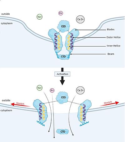
Those mutations could provoke increases in permeability of cations in RBC by different mechanisms. It could induce mechanically activated currents that inactivate more slowly than wild-type currents. They could also affect the inactivation process by either destabilising the inactivated state or stabilising the channel in the open state. As a result, the open to inactivated state equilibrium shifts towards open. Na+ and Ca2+ ion influx consequently increase, and the intracellular K+ concentration decreases in a steady state. The evolution of Piezo1’s function steams from a change in its 3D structure. | Those mutations could provoke increases in permeability of cations in RBC by different mechanisms. It could induce mechanically activated currents that inactivate more slowly than wild-type currents. They could also affect the inactivation process by either destabilising the inactivated state or stabilising the channel in the open state. As a result, the open to inactivated state equilibrium shifts towards open. Na+ and Ca2+ ion influx consequently increase, and the intracellular K+ concentration decreases in a steady state. The evolution of Piezo1’s function steams from a change in its 3D structure. | ||
| - | Lymphatic dysplasia is also a disease linked to loss of function mutations on Piezo1. The lymphatic system is independent from the vascular one, and its role is to transport antigens responsible for the immune response. If the interstitial fluid is not drained correctly back to the blood, it leads to local inflammation. The mutations on Piezo1 inactivate the channel gate and in this case the concentration of calcium is not increased. The protein isn’t sensitive to the pressure anymore. | + | Lymphatic dysplasia <ref name = "Lymphatic dysplasia"> DOI 10.1016/bs.ctm.2017.01.001 </ref> is also a disease linked to loss of function mutations on Piezo1. The lymphatic system is independent from the vascular one, and its role is to transport antigens responsible for the immune response. If the interstitial fluid is not drained correctly back to the blood, it leads to local inflammation. The mutations on Piezo1 inactivate the channel gate and in this case the concentration of calcium is not increased. The protein isn’t sensitive to the pressure anymore. |
== Potential therapeutic target == | == Potential therapeutic target == | ||
Revision as of 15:04, 15 January 2022
| |||||||||||
References
- ↑ 1.0 1.1 1.2 Zhao Q, Wu K, Geng J, Chi S, Wang Y, Zhi P, Zhang M, Xiao B. Ion Permeation and Mechanotransduction Mechanisms of Mechanosensitive Piezo Channels. Neuron. 2016 Mar 16;89(6):1248-1263. doi: 10.1016/j.neuron.2016.01.046. Epub 2016, Feb 25. PMID:26924440 doi:http://dx.doi.org/10.1016/j.neuron.2016.01.046
- ↑ 2.0 2.1 Parpaite T, Coste B. Piezo channels. Curr Biol. 2017 Apr 3;27(7):R250-R252. doi: 10.1016/j.cub.2017.01.048. PMID:28376327 doi:http://dx.doi.org/10.1016/j.cub.2017.01.048
- ↑ 3.0 3.1 3.2 Zhou, Z. (2019). Structural Analysis of Piezo1 Ion Channel Reveals the Relationship between Amino Acid Sequence Mutations and Human Diseases. 139–155. DOI 10.4236/jbm.2019.712012
- ↑ 4.0 4.1 4.2 4.3 4.4 Zhao Q, Zhou H, Chi S, Wang Y, Wang J, Geng J, Wu K, Liu W, Zhang T, Dong MQ, Wang J, Li X, Xiao B. Structure and mechanogating mechanism of the Piezo1 channel. Nature. 2018 Feb 22;554(7693):487-492. doi: 10.1038/nature25743. Epub 2018 Jan, 22. PMID:29469092 doi:http://dx.doi.org/10.1038/nature25743
- ↑ 5.0 5.1 5.2 5.3 Liang X, Howard J. Structural Biology: Piezo Senses Tension through Curvature. Curr Biol. 2018 Apr 23;28(8):R357-R359. doi: 10.1016/j.cub.2018.02.078. PMID:29689211 doi:http://dx.doi.org/10.1016/j.cub.2018.02.078
- ↑ 6.0 6.1 Guo YR, MacKinnon R. Structure-based membrane dome mechanism for Piezo mechanosensitivity. Elife. 2017 Dec 12;6. pii: 33660. doi: 10.7554/eLife.33660. PMID:29231809 doi:http://dx.doi.org/10.7554/eLife.33660
- ↑ 7.0 7.1 7.2 Ge J, Li W, Zhao Q, Li N, Chen M, Zhi P, Li R, Gao N, Xiao B, Yang M. Architecture of the mammalian mechanosensitive Piezo1 channel. Nature. 2015 Nov 5;527(7576):64-9. doi: 10.1038/nature15247. Epub 2015 Sep 21. PMID:26390154 doi:http://dx.doi.org/10.1038/nature15247
- ↑ 8.0 8.1 Saotome K, Murthy SE, Kefauver JM, Whitwam T, Patapoutian A, Ward AB. Structure of the mechanically activated ion channel Piezo1. Nature. 2017 Dec 20. pii: nature25453. doi: 10.1038/nature25453. PMID:29261642 doi:http://dx.doi.org/10.1038/nature25453
- ↑ 9.0 9.1 Lin YC, Guo YR, Miyagi A, Levring J, MacKinnon R, Scheuring S. Force-induced conformational changes in PIEZO1. Nature. 2019 Sep;573(7773):230-234. doi: 10.1038/s41586-019-1499-2. Epub 2019 Aug, 21. PMID:31435018 doi:http://dx.doi.org/10.1038/s41586-019-1499-2
- ↑ 10.0 10.1 10.2 10.3 10.4 Wei L, Mousawi F, Li D, Roger S, Li J, Yang X, Jiang LH. Adenosine Triphosphate Release and P2 Receptor Signaling in Piezo1 Channel-Dependent Mechanoregulation. Front Pharmacol. 2019 Nov 6;10:1304. doi: 10.3389/fphar.2019.01304. eCollection, 2019. PMID:31780935 doi:http://dx.doi.org/10.3389/fphar.2019.01304
- ↑ Li J, Hou B, Tumova S, Muraki K, Bruns A, Ludlow MJ, Sedo A, Hyman AJ, McKeown L, Young RS, Yuldasheva NY, Majeed Y, Wilson LA, Rode B, Bailey MA, Kim HR, Fu Z, Carter DA, Bilton J, Imrie H, Ajuh P, Dear TN, Cubbon RM, Kearney MT, Prasad KR, Evans PC, Ainscough JF, Beech DJ. Piezo1 integration of vascular architecture with physiological force. Nature. 2014 Nov 13;515(7526):279-82. doi: 10.1038/nature13701. Epub 2014 Aug 10. PMID:25119035 doi:http://dx.doi.org/10.1038/nature13701
- ↑ Albuisson J, Murthy SE, Bandell M, Coste B, Louis-Dit-Picard H, Mathur J, Feneant-Thibault M, Tertian G, de Jaureguiberry JP, Syfuss PY, Cahalan S, Garcon L, Toutain F, Simon Rohrlich P, Delaunay J, Picard V, Jeunemaitre X, Patapoutian A. Dehydrated hereditary stomatocytosis linked to gain-of-function mutations in mechanically activated PIEZO1 ion channels. Nat Commun. 2013;4:1884. doi: 10.1038/ncomms2899. PMID:23695678 doi:http://dx.doi.org/10.1038/ncomms2899
- ↑ Yamaguchi Y, Allegrini B, Rapetti-Mauss R, Picard V, Garcon L, Kohl P, Soriani O, Peyronnet R, Guizouarn H. Hereditary Xerocytosis: Differential Behavior of PIEZO1 Mutations in the N-Terminal Extracellular Domain Between Red Blood Cells and HEK Cells. Front Physiol. 2021 Oct 18;12:736585. doi: 10.3389/fphys.2021.736585. eCollection, 2021. PMID:34737711 doi:http://dx.doi.org/10.3389/fphys.2021.736585
- ↑ Alper SL. Genetic Diseases of PIEZO1 and PIEZO2 Dysfunction. Curr Top Membr. 2017;79:97-134. doi: 10.1016/bs.ctm.2017.01.001. Epub 2017 Feb, 28. PMID:28728825 doi:http://dx.doi.org/10.1016/bs.ctm.2017.01.001
- ↑ Shinge SAU, Zhang D, Achu Muluh T, Nie Y, Yu F. Mechanosensitive Piezo1 Channel Evoked-Mechanical Signals in Atherosclerosis. J Inflamm Res. 2021 Jul 27;14:3621-3636. doi: 10.2147/JIR.S319789. eCollection, 2021. PMID:34349540 doi:http://dx.doi.org/10.2147/JIR.S319789
- ↑ Velasco-Estevez M, Gadalla KKE, Linan-Barba N, Cobb S, Dev KK, Sheridan GK. Inhibition of Piezo1 attenuates demyelination in the central nervous system. Glia. 2020 Feb;68(2):356-375. doi: 10.1002/glia.23722. Epub 2019 Oct 9. PMID:31596529 doi:http://dx.doi.org/10.1002/glia.23722
- ↑ Chubinskiy-Nadezhdin VI, Vasileva VY, Vassilieva IO, Sudarikova AV, Morachevskaya EA, Negulyaev YA. Agonist-induced Piezo1 activation suppresses migration of transformed fibroblasts. Biochem Biophys Res Commun. 2019 Jun 18;514(1):173-179. doi:, 10.1016/j.bbrc.2019.04.139. Epub 2019 Apr 24. PMID:31029419 doi:http://dx.doi.org/10.1016/j.bbrc.2019.04.139
- ↑ Liu S, Pan X, Cheng W, Deng B, He Y, Zhang L, Ning Y, Li J. Tubeimoside I Antagonizes Yoda1-Evoked Piezo1 Channel Activation. Front Pharmacol. 2020 May 25;11:768. doi: 10.3389/fphar.2020.00768. eCollection, 2020. PMID:32523536 doi:http://dx.doi.org/10.3389/fphar.2020.00768

