User:George G. Papadeas/Sandbox VKOR
From Proteopedia
(Difference between revisions)
| Line 6: | Line 6: | ||
== Introduction== | == Introduction== | ||
=== Biological Role === | === Biological Role === | ||
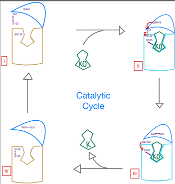
| - | <scene name='90/906893/Vkor_structure/1'>Vitamin K epoxide reductase</scene> (VKOR) is a reducing enzyme composed of 4-helices that spans the endoplasmic reticulum as a transmembrane protein <ref>DOI 10.1126</ref>. Its enzymatic role is reducing <scene name='90/906893/Vkor_with_ko/1'>vitamin K epoxide</scene> (KO) to Vitamin K Hydroquinone (KH2) <ref>DOI 10.1021</ref> (Figure 1). The mechanism first occurs through the binding KO and using two cysteine residues to reduce KO into [https://en.wikipedia.org/wiki/Vitamin_K Vitamin K]. Then, a second pair of cysteine residues will reduce Vitamin K into the final product, KH2 (Figure 1). One of VKORs primary roles is to assist in blood coagulation through this KH2 regeneration mechanism.[[Image:VKOR_mechanism_2D.png|400 px|right|thumb|Figure 1. Mechanism of KO oxidation into KH2.]] With Vitamin K as a cofactor, the [https://www.britannica.com/science/bleeding/The-extrinsic-pathway-of-blood-coagulation#ref64617 γ-carboxylase] enzyme will enact post-translational modification on KH2, oxidizing it back to KO. The oxidation of KH2 by γ-carboxylase is coupled with the carboxylation of a glutamate residue to form γ-carboxyglutamate. The coupling of this oxidation and carboxylation will activate several clotting factors in the coagulation cascade. | + | <scene name='90/906893/Vkor_structure/1'>Vitamin K epoxide reductase</scene> (VKOR) is a reducing enzyme composed of 4-helices that spans the endoplasmic reticulum as a transmembrane protein <ref>DOI 10.1126</ref>. Its enzymatic role is reducing <scene name='90/906893/Vkor_with_ko/1'>vitamin K epoxide</scene> (KO) to Vitamin K Hydroquinone (KH2) <ref>DOI 10.1021</ref> (Figure 1). The mechanism first occurs through the binding KO and using two cysteine residues to reduce KO into [https://en.wikipedia.org/wiki/Vitamin_K Vitamin K]. Then, a second pair of cysteine residues will reduce Vitamin K into the final product, KH2 (Figure 1). One of VKORs primary roles is to assist in blood coagulation through this KH2 regeneration mechanism.[[Image:VKOR_mechanism_2D.png|400 px|right|thumb|Figure 1. Mechanism of KO oxidation into KH2.]] With Vitamin K as a cofactor, the [https://www.britannica.com/science/bleeding/The-extrinsic-pathway-of-blood-coagulation#ref64617 γ-carboxylase] enzyme will enact post-translational modification on KH2, oxidizing it back to KO. The oxidation of KH2 by γ-carboxylase is coupled with the carboxylation of a glutamate residue to form γ-carboxyglutamate. The coupling of this oxidation and carboxylation will activate several clotting factors in the coagulation cascade. |
| + | |||
=== Author's Notes === | === Author's Notes === | ||
Structural characterization of VKOR has been difficult due to its in vitro instability. Recently, a series of atomic structures have been determined utilizing anticoagulant stabilization and VKOR-like [https://pubmed.ncbi.nlm.nih.gov/33154105/ homologs]. Crystal structures of VKOR were captured with a bound substrate (KO) or vitamin K antagonist (VKA) (PDB Codes: Table 1)<ref>DOI 10.1126</ref>. VKA substrates utilized were anticoagulants, namely [https://en.wikipedia.org/wiki/Warfarin Warfarin], [https://en.wikipedia.org/wiki/Brodifacoum Brodifacoum], [https://en.wikipedia.org/wiki/Phenindione Phenindione], and [https://en.wikipedia.org/wiki/Chlorophacinone Chlorophacinone]. Second, VKOR-like homologs were utilized to aid in structure classification. Homologs refer to specific cysteine residues that have been mutated to serine to facilitate capturing a stable conformation state. Homologs were mainly isolated from human VKOR with some isolated from the pufferfish ''Takifugu rubripes''. Furthermore, all of the structures used have been processed to remove a beta barrel at the south end of VKOR that served no purpose in function of the enzyme. This also allowed for the residue numbering to be reassigned and more closely replicate the human VKOR. | Structural characterization of VKOR has been difficult due to its in vitro instability. Recently, a series of atomic structures have been determined utilizing anticoagulant stabilization and VKOR-like [https://pubmed.ncbi.nlm.nih.gov/33154105/ homologs]. Crystal structures of VKOR were captured with a bound substrate (KO) or vitamin K antagonist (VKA) (PDB Codes: Table 1)<ref>DOI 10.1126</ref>. VKA substrates utilized were anticoagulants, namely [https://en.wikipedia.org/wiki/Warfarin Warfarin], [https://en.wikipedia.org/wiki/Brodifacoum Brodifacoum], [https://en.wikipedia.org/wiki/Phenindione Phenindione], and [https://en.wikipedia.org/wiki/Chlorophacinone Chlorophacinone]. Second, VKOR-like homologs were utilized to aid in structure classification. Homologs refer to specific cysteine residues that have been mutated to serine to facilitate capturing a stable conformation state. Homologs were mainly isolated from human VKOR with some isolated from the pufferfish ''Takifugu rubripes''. Furthermore, all of the structures used have been processed to remove a beta barrel at the south end of VKOR that served no purpose in function of the enzyme. This also allowed for the residue numbering to be reassigned and more closely replicate the human VKOR. | ||
| Line 31: | Line 32: | ||
== Disease and Treatment == | == Disease and Treatment == | ||
=== Afflictions === | === Afflictions === | ||
| + | |||
=== Inhibition === | === Inhibition === | ||
| + | [[Image:Warfarin.png |400 px| right| thumb]] | ||
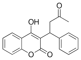
The most inexpensive and common way to treat blood clotting is through the VKOR inhibitor, <scene name='90/906893/Vkor_with_warfarin_bound/2'>Warfarin</scene>. [https://en.wikipedia.org/wiki/Warfarin Warfarin] is able to do so by outcompeting KO. It will enter the binding pocket of VKOR, creating strong hydrogen bonds with the active site. | The most inexpensive and common way to treat blood clotting is through the VKOR inhibitor, <scene name='90/906893/Vkor_with_warfarin_bound/2'>Warfarin</scene>. [https://en.wikipedia.org/wiki/Warfarin Warfarin] is able to do so by outcompeting KO. It will enter the binding pocket of VKOR, creating strong hydrogen bonds with the active site. | ||
| + | |||
=== Mutations === | === Mutations === | ||
Some key <scene name='90/906893/Active_site_mutations/2'>mutations</scene> that can be detrimental to the VKOR structure are mutations of the <scene name='90/906893/Active_site/4'>active site</scene>. The two main residues, N80 and Y139, can be mutated to A80 and F139 creating a decrease in recognition and stabilization. | Some key <scene name='90/906893/Active_site_mutations/2'>mutations</scene> that can be detrimental to the VKOR structure are mutations of the <scene name='90/906893/Active_site/4'>active site</scene>. The two main residues, N80 and Y139, can be mutated to A80 and F139 creating a decrease in recognition and stabilization. | ||
| Line 38: | Line 42: | ||
This is a sample scene created with SAT to <scene name="/12/3456/Sample/1">color</scene> by Group, and another to make <scene name="/12/3456/Sample/2">a transparent representation</scene> of the protein. You can make your own scenes on SAT starting from scratch or loading and editing one of these sample scenes. | This is a sample scene created with SAT to <scene name="/12/3456/Sample/1">color</scene> by Group, and another to make <scene name="/12/3456/Sample/2">a transparent representation</scene> of the protein. You can make your own scenes on SAT starting from scratch or loading and editing one of these sample scenes. | ||
| - | + | ||
</StructureSection> | </StructureSection> | ||
Revision as of 02:39, 18 April 2022
VKOR
| |||||||||||
References
1. DJin, Da-Yun, Tie, Jian-Ke, and Stafford, Darrel W. "The Conversion of Vitamin K Epoxide to Vitamin K Quinone and Vitamin K Quinone to Vitamin K Hydroquinone Uses the Same Active Site Cysteines." Biochemistry 2007 46 (24), 7279-7283 [1].
2. Li, Weikai et al. “Structure of a bacterial homologue of vitamin K epoxide reductase.” Nature vol. 463,7280 (2010): 507-12. doi:10.1038/nature08720.
3. Liu S, Li S, Shen G, Sukumar N, Krezel AM, Li W. Structural basis of antagonizing the vitamin K catalytic cycle for anticoagulation. Science. 2021 Jan 1;371(6524):eabc5667. doi: 10.1126/science.abc5667. Epub 2020 Nov 5. PMID: 33154105; PMCID: PMC7946407.
- ↑ Hanson, R. M., Prilusky, J., Renjian, Z., Nakane, T. and Sussman, J. L. (2013), JSmol and the Next-Generation Web-Based Representation of 3D Molecular Structure as Applied to Proteopedia. Isr. J. Chem., 53:207-216. doi:http://dx.doi.org/10.1002/ijch.201300024
- ↑ Herraez A. Biomolecules in the computer: Jmol to the rescue. Biochem Mol Biol Educ. 2006 Jul;34(4):255-61. doi: 10.1002/bmb.2006.494034042644. PMID:21638687 doi:10.1002/bmb.2006.494034042644
- ↑ Unknown PubmedID 10.1126
- ↑ Unknown PubmedID 10.1021
- ↑ Unknown PubmedID 10.1126


