We apologize for Proteopedia being slow to respond. For the past two years, a new implementation of Proteopedia has been being built. Soon, it will replace this 18-year old system. All existing content will be moved to the new system at a date that will be announced here.
Sandbox Reserved 1710
From Proteopedia
(Difference between revisions)
| Line 6: | Line 6: | ||
== Introduction == | == Introduction == | ||
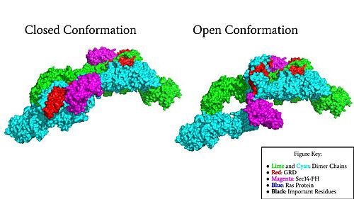
[[Image:Neurofibromin Surface w Labels.jpg|500 px|right|thumb|Figure 1: Surface Rendering of Neurofibromin in its Open (7PGT) and Closed (7PGR) Conformation.]] | [[Image:Neurofibromin Surface w Labels.jpg|500 px|right|thumb|Figure 1: Surface Rendering of Neurofibromin in its Open (7PGT) and Closed (7PGR) Conformation.]] | ||
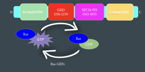
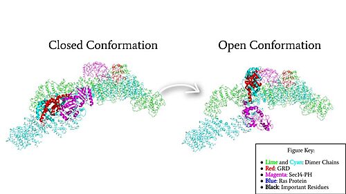
| - | Neurofibromin functions as a tumor suppressor protein by associating with [https://proteopedia.org/wiki/index.php/Ras Ras], a protein that | + | Neurofibromin functions as a tumor suppressor protein by associating with [https://proteopedia.org/wiki/index.php/Ras Ras], a protein that signals for [https://en.wikipedia.org/wiki/Cell_proliferation cell proliferation] and growth. It has two conformations, the open, active conformation and the closed, inactive conformation (Figure 1). <ref name="Trovó-Marqui">PMID:16813595</ref> The change in conformation of Neurofibromin from open to closed regulates Ras activity, turning it off, thus stopping cell growth from continuing in excess. Neurofibromin is a [https://en.wikipedia.org/wiki/Dimer_(chemistry) dimer] made up of two identical [https://en.wikipedia.org/wiki/Monomer monomers] consisting of the Sec 14-PH domain and the GRD (Gap-related domain). The orientation of these domains is important for the interaction and binding of Ras to the Arginine finger in Neurofibromin, a key interaction in the regulation of Ras activity. Neurofibromin in its open conformation allows for the Ras association, unlike the closed conformation which is unable to associate with Ras and turn it off. <ref name="Naschberger">PMID:34707296</ref> The closed conformation is stabilized by a triad consisting of Cys 1032, His 1558, and His 1576 and a Zinc atom. The molecular structure and detail of Neurofibromin have been determined by [https://en.wikipedia.org/wiki/Cryogenic_electron_microscopy Cryo-Electron Microscopy]. Additionally, the structure of Neurofibromin isoform 2 revealed different functional states for Neurofibromin. <ref name="Naschberger">PMID:34707296</ref> Neurofibromin is encoded by the [https://en.wikipedia.org/wiki/Neurofibromin_1 NF1 gene], which is located on chromosome 17. Mutations in the NF1 gene are associated with diseases including Neurofibromatosis Type 1, [https://en.wikipedia.org/wiki/Neurofibroma Plexiform Neurofibromas], and cancers including glioblastoma, neuroblastoma, lung, ovarian, and breast cancer. <ref name="Lupton">PMID: 34887559 </ref> <ref name="Ratner">PMID:25877329</ref> <ref name="Abramowicz">PMID:25182393</ref> |
== Function == | == Function == | ||
Revision as of 02:20, 19 April 2022
| This Sandbox is Reserved from February 28 through September 1, 2022 for use in the course CH462 Biochemistry II taught by R. Jeremy Johnson at the Butler University, Indianapolis, USA. This reservation includes Sandbox Reserved 1700 through Sandbox Reserved 1729. |
To get started:
More help: Help:Editing |
Human Neurofibromin - The Tumor Suppressor Gene
| |||||||||||
References
- ↑ 1.0 1.1 1.2 Trovo-Marqui AB, Tajara EH. Neurofibromin: a general outlook. Clin Genet. 2006 Jul;70(1):1-13. doi: 10.1111/j.1399-0004.2006.00639.x. PMID:16813595 doi:http://dx.doi.org/10.1111/j.1399-0004.2006.00639.x
- ↑ 2.0 2.1 2.2 2.3 2.4 2.5 2.6 Naschberger A, Baradaran R, Rupp B, Carroni M. The structure of neurofibromin isoform 2 reveals different functional states. Nature. 2021 Nov;599(7884):315-319. doi: 10.1038/s41586-021-04024-x. Epub 2021, Oct 27. PMID:34707296 doi:http://dx.doi.org/10.1038/s41586-021-04024-x
- ↑ 3.0 3.1 Lupton CJ, Bayly-Jones C, D'Andrea L, Huang C, Schittenhelm RB, Venugopal H, Whisstock JC, Halls ML, Ellisdon AM. The cryo-EM structure of the human neurofibromin dimer reveals the molecular basis for neurofibromatosis type 1. Nat Struct Mol Biol. 2021 Dec;28(12):982-988. doi: 10.1038/s41594-021-00687-2., Epub 2021 Dec 9. PMID:34887559 doi:http://dx.doi.org/10.1038/s41594-021-00687-2
- ↑ 4.0 4.1 4.2 Ratner N, Miller SJ. A RASopathy gene commonly mutated in cancer: the neurofibromatosis type 1 tumour suppressor. Nat Rev Cancer. 2015 May;15(5):290-301. doi: 10.1038/nrc3911. Epub 2015 Apr 16. PMID:25877329 doi:http://dx.doi.org/10.1038/nrc3911
- ↑ 5.0 5.1 5.2 Abramowicz A, Gos M. Neurofibromin in neurofibromatosis type 1 - mutations in NF1gene as a cause of disease. Dev Period Med. 2014 Jul-Sep;18(3):297-306. PMID:25182393
- ↑ Bergoug M, Doudeau M, Godin F, Mosrin C, Vallee B, Benedetti H. Neurofibromin Structure, Functions and Regulation. Cells. 2020 Oct 27;9(11). pii: cells9112365. doi: 10.3390/cells9112365. PMID:33121128 doi:http://dx.doi.org/10.3390/cells9112365


